Transcription of LOGIQ F8 - media2.lojer.com
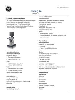
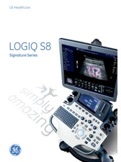
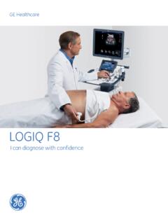
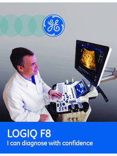
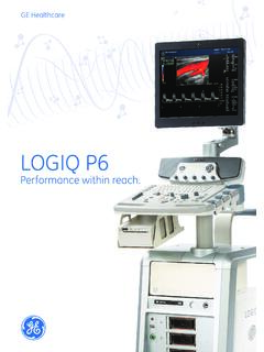
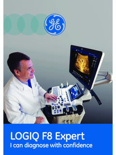
1 LOGIQ F8 Data SheetGE HealthcareThe LOGIQ * F8 is the multipurpose ultrasound imaging system designed for Abdominal, Obstetrical, Gynecological, Small Parts, Musculoskeletal, Vascular/Peripheral Vascular, Urological, Pediatric, Transcranial and Cardiac specificationsDimensions and WeightHeight with 19'' LCD Max 1495mm ( in) Min 1410mm ( in) Width Keyboard: 500 mm ( in) Caster: 720 mm ( in)Depth Maximum: 810 mm ( in) Caster: 800 mm ( in)Weight (no Peripherals)58 kgElectrical PowerVoltage 100-240 VACF requency 50/60 HzPower consumption maximum of 400 VA with peripheralsConsole DesignMax 4 active probe portsIntegrated HDD Integrated SpeakersProbe HoldersGel HolderFront and Rear HandlesProbe Cable TrayUser interfaceOperator KeyboardErgonomic full size keyboard8 TGC ( mm) LCD touch screenMonitor19'' ( mm)
2 High-resolution LCDA rticulating monitor armSystem overviewApplicationsAbdominal ObstetricalGynecological Small PartsMusculoskeletalVascular/Peripheral Vascular UrologicalPediatric Transcranial CardiacScanning MethodsElectronic ConvexElectronic LinearElectronic Micro ConvexElectronic SectorReal Time 4D Volume SweepTransducer TypesConvex ArrayLinear ArrayMicroconvex ArraySector Phased ArrayVolume Probes (4D)Operating ModesB-ModeCoded Phase Inversion Harmonic ImagingM-ModeColor M-ModeColor Flow Mode (CFM)Power Doppler Imaging (PDI)Directional PDIPW Doppler with High PRFA natomical M-Mode (Option)CW Doppler Mode (Option)LOGIQView (Option)TVI Mode (Option)3D/4D Volume Modes (Option)System overview (continued)System Standard FeaturesAO (Automatic Optimization) CrossXBeam*SRI-HD (High Definition Speckle Reduction Imaging)
3 B-SteerCoded Phase Inversion Harmonic ImagingVirtual ConvexPatient Information DatabaseImage Archive on integrated HDDRaw Data AnalysisScan AssistantScan CoachReal-Time Automatic Doppler CalculationsOB CalculationsFetal TrendingMultigestational CalculationsHip Dysplasia CalculationsGynecological CalculationsVascular CalculationsUrological CalculationsRenal CalculationsCardiac CalculationsRemote capability: InSite* ExCOn-Board Reporting PackageMPEGVueNetwork StorageSystem OptionsElastographyLOGIQViewAuto IMTCW DopplerAnatomical M-ModeTissue Velocity Imaging (TVI)
4 With Q-AnalysisEasy 3 DStatic 3D/ Realtime 4D DICOM ConnectivityExtra Probe HolderPaper TrayPeripheral OptionsFixture Kit for Digital UP-D711 Thermal PrinterDigital UP-D711 Thermal PrinterDigital UP-D25 Color Thermal PrinterDigital UP-D897 BW Thermal PrinterHP office jet 100 Mobile Printer 1-Pedal Type Footswitch Whanam FSU-1000 Footswitch MKF 2-MED USB GP26 SanDisk USB Stick 4G1TB Mobile USB HDDUSB LampUSB ECG Kits (AHA/IEC)Display ModesLive and Stored Display Format: full size and split screen both with thumbnails for still and CineReview Image Format.
5 4x4 and thumbnails for still and CineSimultaneous CapabilityB or CrossXBeam/PWB or CrossXBeam/CFM or PDIB/MB/CrossXBeamReal-Time Triplex Mode (B or CrossXBeam + CFM or PDI/PW)Selectable Alternating ModesB or CrossXBeam/PWB or CrossXBeam + CFM (PDI)/PW(CW(Option))B/CW (Option)Multi-image (split/quad screen)Live and/or FrozenB or CrossXBeam + B or CrossXBeam/CFM or PDII ndependent Cine PlaybackTime Line DisplayIndependent Dual B or CrossXBeam/PW DisplayCWDisplay Formats Top/Bottom Selectable Format Side/Side Selectable FormatVirtual ConvexTimeline OnlySystem overview (continued)Display AnnotationPatient Name: First , Last Patient IDOther IDAge, Sex and Birth DateHospital NameDate format: 3 Types Selectable MM/DD/YY DD/MM/YY YY/MM/DDTime format.
6 2 Types Selectable 24 hours 12 hoursGestational Age LMP GA EDD BBTD isplayed Acoustic Output TIS: Thermal Index Soft Tissue TIC: Thermal Index Cranial (Bone) TIB: Thermal Index Bone MI: Mechanical Index% of Maximum Power OutputProbe NameMap NamesProbe OrientationDepth Scale MarkerLateral Scale MarkerFocal Zone MarkersImage DepthZoom DepthB-ModeGainDynamic RangeImaging FrequencyFrame AveragingAcoustic Frame RateGray MapSRI-HDM-ModeGainDynamic RangeTime ScaleDoppler ModeGainAngleSample Volume Depth and WidthDisplay Annotation (continued)Wall FilterVelocity and/or Frequency ScaleSpectrum InversionTime ScalePRFD oppler FrequencyColor Flow ModeLine DensityFrame AveragingPacket SizeColor Scale.
7 2 Types Power Directional PDIC olor Velocity Range and BaselineColor Threshold MarkerColor GainPDII nversionDoppler FrequencyTGC CurveCine Gage, Image Number/Frame NumberBody Pattern: Multiple human Application NameMeasurement ResultsOperator MessageBiopsy Guide Line and ZoneHeart RateGeneral system parametersSystem SetupPre-Programmable CategoriesUser Programmable Preset CapabilityFactory Default Preset DataLanguages: English, Latin American Spanish, French, German, Italian, Brazilian Portuguese, Chinese (Simplified), Swedish, Russian, Norwegian, Danish, Dutch, Finnish, JapaneseOB Report Formats Including Tokyo Univ.
8 , Osaka Univ., USA, Europe, and ASUMUser Defined AnnotationsBody PatternsCustomized Comment Home PositionCINE Memory/Image Memory128 MB of Cine MemorySelectable Cine Sequence for Cine ReviewProspective Cine MarkMeasurements/Calculations and Annotations on Cine PlaybackScrolling Timeline MemoryDual Image Cine DisplayQuad Image Cine DisplayCine Gauge and Cine Image Number DisplayCine Review LoopCine Review SpeedConnectivity & DICOME thernet Network ConnectionDICOM (Optional)VerifyPrintStoreModality WorklistStorage CommitmentModality Performed Procedure Step (MPPS)Query/RetrieveStructured Reporting Template which can be compared to vascular and OB standard valuesRemote Capability InSite ExCScanning ParametersDisplayed Imaging Depth: 0 33 cmMinimum Depth of Field: 0 2 cm (Zoom) (probe dependent)Maximum Depth of Field: 0 33 cm (probe dependent)Continuous Dynamic Receive Focus/Continuous Dynamic Receive ApertureAdjustable Dynamic RangeAdjustable Field of View (FOV)Image Reverse.
9 Right/LeftImage Rotation of 0 , 180 B-Mode Adjustable:Acoustic PowerGainDynamic RangeFrame AveragingGray Scale MapFrequencyLine DensityScanning Size (FOV or Angle depending on the probe, see probe specifications)B ColorizationRejectSuppressionSRI-HDEdge EnhanceImage StorageOn-Board Database of Patient InformationStorage FormatsDICOM compressed/uncompressed, single/multiframe, with/without Raw DataExport JPEG, JPEG2000, WMV (MPEG 4) and AVI formatsStorage DevicesUSB Memory StickDVD-RW StorageHDD Image StorageGeneral system parameters (continued)M-ModeAdjustable.
10 Acoustic PowerGainDynamic RangeGray Scale MapSweep SpeedM ColorizationM Display FormatRejectionAnatomical M-Mode (Option)M-Mode cursor adjustable at any planeCan be activated from a Cine loop from a live or stored imageM and A CapabilityAvailable with Color Flow ModePulse Wave Doppler ModeAdjustable:Acoustic PowerGainGray Scale MapTransmit FrequencyWall FilterPW ColorizationVelocity Scale RangeSweep SpeedSample Volume DepthAngle CorrectionSpectrum InversionTrace MethodBaseline ShiftDoppler Auto TraceCompressionTrace DirectionTrace SensitivityColor Flow ModeAdjustable:Acoustic PowerColor Maps, including velocity-variance mapsGainVelocity Scale RangeWall FilterPacket SizeLine DensitySpatial FilterSteering AngleBaseline ShiftFrame AverageThresholdAccumulation ModeSample Volume ControlFlash SuppressionPower Doppler ImagingAdjustable.