Transcription of LOGIQ S8 R2 Specifications - Davis Medical Electronics
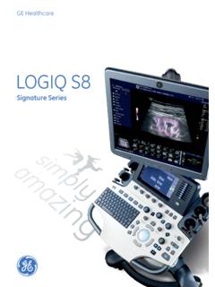
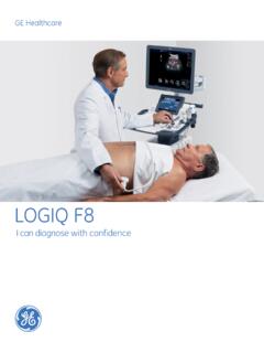
1 ge healthcare Chalfont St. Giles Buckinghamshire, UK DOC1487369: LOGIQ S8 R2 Specification Sheet LOGIQ S8 Product Description The LOGIQ * S8 is our premium multi-purpose ultrasound imaging system designed for abdominal, vascular, breast, cardiac, small parts, obstetrics, gynecology, neonatal, pediatrics, urology and transcranial applications. DOC1487369: LOGIQ S8 R2 Specification Sheet Page 2 of 14 General Specifications Dimensions and Weight Height Maximum: 1750mm, Minimum: 1150mm, Width Keyboard: 500mm, in Caster: 620mm, in Depth Maximum: 880mm, in Caster: 790mm, in Weight 85 kg, lbs Electrical Power Voltage: 100-120 Vac or 220-240 Vac Frequency: 50/60 Hz Power consumption maximum of 900VA with peripherals Console Design 4 Active probe ports and 1 parking Integrated HDD (at least 500 GB) Integrated DVD multi drive On-board storage of thermal printer Integrated speakers Locking mechanism that provides rolling lock and caster swivel lock Integrated cable management Front and rear handles Easily removable air filters User Interface Operator Keyboard Operating keyboard adjustable in two dimensions.
2 Height Rotation Backlit alphanumeric keyboard Ergonomic hard key layout Interactive back-lighting Integrated recording keys for remote control of up to 6 peripheral or DICOM** devices Integrated gel warmer Touch Screen Wide 9 high-resolution, color, touch, LCD screen Interactive dynamic software menu Brightness adjustment User-configurable layout LCD Monitor 19 high-resolution LCD LCD translation (independent of console): 660 mm horizontal (end to end) 135 mm vertical (end to end) 90 swivel Fold-down and lock mechanism for transportation Brightness and contrast adjustment Resolution: 1280 X 1024 Horizontal/Vertical viewing angle of +/- 170 System Overview Applications Abdominal Obstetrical Gynecological Breast Small parts Vascular /Peripheral Transcranial Pediatrics and neonatal Musculoskeletal Urological Cardiac Operating Modes B-Mode M-Mode Color Flow Mode (CFM) TVI (Option) B-Flow*/B-Flow Color (Option) Extended Field of View ( LOGIQ View, Option) Power Doppler Imaging (PDI) PW Doppler CW Doppler (Option) Volume Modes (3D/4D) Static 3D Real Time 4D (option) Anatomical M-Mode Curved Anatomical M-Mode B Steer+ (Option) Coded Contrast Imaging (Option) Elastography (Option) Scanning Methods Electronic Sector Electronic Convex Electronic Linear Mechanical Volume Sweep Transducer Types Sector Phased Array Convex Array Micro convex Array Linear Array Matrix Array Volume Probes (4D)
3 Convex Array Micro convex Array Split Crystal System Standard Features Advanced user interface with high resolution wide 9 LCD touch panel Automatic Optimization CrossXBeam* Speckle Reduction Imaging (SRI-HD) Fine Angle Steer Coded Harmonic Imaging Virtual Convex Patient information database Image Archive on integrated CD/DVD and hard drive 3D Raw Data Analysis Real-time automatic Doppler calcs OB Calcs Fetal Trending Multigestational Calcs Hip Dysplasia Calcs Gynecological Calcs Vascular Calcs Urological Calcs Renal Calcs Cardiac Calcs InSite*ExC capability On-board electronic documentation Peripheral Options Integrated options for: Digital BW thermal printer DVD video recorder Digital color thermal printer Digital A6 color thermal printer External USB printer connection DVI-D output available for compatible devices Foot Switch, with programmable functionality Video Converter Display Modes Live and Stored Display Format: Full size and split screen - both w/ thumbnails.
4 For still and Cine Review Image Format: 4x4 and "thumbnails". for still and Cine Simultaneous Capability B or CrossXBeam /PW B or CrossXBeam /CFM or PDI DOC1487369: LOGIQ S8 R2 Specification Sheet Page 3 of 14 B/M B/CrossXBeam Real-time Triplex Mode (B or CrossXBeam + CFM or PDI/PW or CW(Option)) Selectable alternating Modes B or CrossXBeam /PW B or CrossXBeam + CFM (PDI)/PW(CW(Option)) B/CW (Option) Multi-image (split/quad screen) Live and/or frozen B or CrossXBeam + B or CrossXBeam /CFM or PDI PW/M Independent Cine playback Time line display Independent Dual B or CrossXBeam /PW Display CW Display Formats Top/ Bottom selectable format Side/Side selectable format Timeline only Virtual Convex Display Annotation Patient Name: First, Last and Middle Patient ID 2nd Patient ID Age, Sex and Birth Date Hospital Name Date format.
5 3 types selectable MM/DD/YY, DD/MM/YY, YY/MM/DD Time format: 2 types selectable: 24 hours, 12 hours Gestational Age from LMP/EDD/GA/BBT Probe Name Map names Probe Orientation Depth Scale Marker Lateral Scale Marker Focal Zone Markers Image Depth Zoom Depth B-Mode Gain Dynamic Range Imaging Frequency Frame Averaging Gray Map SRI-HD M-Mode Gain Dynamic Range Time Scale Doppler Mode Gain Angle Sample Volume Depth and Width Wall Filter Velocity and/or Frequency Scale Spectrum Inversion Time Scale PRF Doppler Frequency Color Flow Mode Line Density Frame Averaging Packet Size Color Scale: 3 types Power, Directional PDI, and Symmetrical Velocity Imaging Color Velocity Range and Baseline Color Threshold Marker Color Gain PDI Inversion Doppler Frequency TGC Curve Acoustic Frame Rate Cine Gage, Image Number / Frame Number Body Pattern: Multiple human and animal types Application Name Measurement Results Operator Message Displayed Acoustic Output TIS: Thermal Index Soft Tissue TIC: Thermal Index Cranial (Bone) TIB: Thermal Index Bone MI: Mechanical Index % of Maximum Power output Biopsy Guide Line and Zone Heart Rate General System Parameters System Setup Pre-programmable Categories User Programmable Preset Capability Factory Default Preset Data Languages.
6 English, French, German, Spanish, Italian, Portuguese, Russian, Greek, Swedish, Danish, Dutch, Finnish, Norwegian, Japanese (message only), Chinese (message only) OB Report Formats including Tokyo Univ., Osaka Univ., USA, Europe, and ASUM User Defined Annotations Body Patterns Customized Comment Home Position User Manual available on board through Help (F1) User Manual and Service Manual are included on CD with each system. A printed manual is available upon request. Cine Memory/Image Memory 776 MB of Cine Memory Selectable Cine Sequence for Cine Review Prospective Cine Mark Measurements/ Calculations and Annotations on Cine Playback Scrolling timeline memory Dual Image Cine Display Quad Image Cine Display Cine Gauge and Cine Image Number Display Cine Review Loop Cine Review Speed: 11 steps (11, 13, 14, 17, 22, 25, 31, 48, 100, 200, 400%) Image Storage On-board database of patient information from past exams Storage Formats: DICOM compressed/ uncompressed, single/ multi frame, with/ without Raw Data Export JPEG, JPEG2000, WMV (MPEG 4) and AVI formats Storage Devices: USB Memory Stick: 64MB to 4GB (for exporting individual images/clips) CD-RW storage: 700MB DVD storage: -R ( ) Hard Drive Image Storage.
7 ~112GB Compare previous exam images with current exam images Reload of archived data sets Network Storage support for Import, Export, DICOM Read, SaveAs, SaveAs Image, Report SaveAs, MPEGVue DOC1487369: LOGIQ S8 R2 Specification Sheet Page 4 of 14 Connectivity and DICOM Ethernet network connection DICOM (Option) Verify Print Store Modality Worklist Storage Commitment Modality Performed Procedure Step (MPPS) Media Exchange Off network / mobile storage queue Query / Retrieve Public SR Template Structured Reporting compatible with vascular and OB standard InSite ExC capability Physiological Input Panel (Option) Physiological Input ECG, 2 lead Dual R-Trigger Pre-settable ECG R Delay Time Pre-settable ECG Position Adjustable ECG Gain Control Automatic Heart Rate Display Report Writer (Option)
8 On-board reporting package automates report writing Formats various exam results into a report suitable for printing or reviewing on a standard PC Exam result reports can include patient info, exam info, measurements, calculations, images, comments and physician diagnosis Standard templates provided Customizable templates Scanning Parameters Digital Beamformer 478,405 Channels Frame Rate: 2399Hz Maximum Displayed Imaging Depth: 0 33 cm Minimum Depth of Field: 0 2 cm (Zoom) (probe dependent) Maximum Depth of Field: 0 33 cm (probe dependent) Continuous Dynamic Receive Focus / Continuous Dynamic Receive Aperture 274dB Dynamic Range System frequency range: 1-13 MHz 256 shades of gray Adjustable Field of View (FOV) Image Reverse: Right/ Left Image Rotation of 0 , 180 Digital B-Mode Adjustable: Acoustic Power: 0 - 100%, 25 steps Gain: 0 90 dB range, 1dB steps Dynamic Range: 36 96 dB, 16 steps Frame Averaging: up to 8 steps Gray Scale Map: 10 maps Frequency: up to 6 steps Speed of Sound (probe, application dependent) Line Density: up to 5 steps Scanning Size (FOV or Angle - depending on the probe, see probe Specifications ) B Colorization: 9 maps Reject: 6 steps Suppression: up to 6 steps SRI-HD: 0 5, 6 steps Edge Enhance: 7 steps Digital M-Mode Adjustable: Acoustic Power: 0 - 100%, 25 steps Gain: -20 20 dB (delta from B) Dynamic Range : 36 96 dB, 16 steps Gray Scale Map: 10 maps Frequency: up to 6 steps Sweep Speed: 0 7, 8 steps M Colorization: 9 maps M Display Format: V-1/3B, V-1/2B, V-2/3B, H-1/2B, H-1/4B, TL Only Rejection.
9 6 steps Anatomical M-Mode M-mode cursor adjustable at any plane Can be activated from a Cine loop from a live or stored image M and A capability Available with Color Flow Mode Curved Anatomical M-Mode Digital Spectral Doppler Mode Adjustable: Acoustic Power: 0-100%, 25 steps Gain: 0 85dB range, 1dB steps Gray Scale Map: 8 maps Transmit Frequency: Up to 5 steps Wall Filter: 18 settings, Min: 5 Hz, Max: 3,339Hz PW Colorization: 6 maps Velocity Scale Range: Sweep Speed: 0 7, 8 steps Sample Volume Length: 1 16mm, 12 steps Angle Correction: -90 - 90 degrees, 1 degree steps Steered Linear: 0 30 degrees Spectrum Inversion Trace Method Baseline Shift: 5 95%, 11 steps Doppler Auto Trace Compression: , 9 steps Trace Direction Trace Sensitivity Digital Color Flow Mode Adjustable: Acoustic Power: 0 - 100%, 25 steps Color Maps, including velocity-variance maps: 15 maps Gain: 0 40dB, 81 steps Velocity Scale Range: 2 370cm/s Wall Filter: 0 3, 4 steps Packet Size: 5 24, 9 steps Line Density: 5 steps Spatial Filter: 6 steps Steering Angle: 0 20 degrees Baseline Shift: 0 100%, 11 steps Frame Average: 0 6, 7 steps Threshold: 0 100%, 11 steps Accumulation mode: 7 levels Sample Volume Control Flash Suppression Digital Power Doppler Imaging Adjustable: Acoustic Power: 0 - 100%, 25 steps Color Map: 16 maps Gain: 0 40 dB, 81 steps Wall Filter: 0 3, 4 steps Packet Size: 5 24, 9 steps DOC1487369: LOGIQ S8 R2 Specification Sheet Page 5 of 14 Line Density: 5 steps Spatial Filter: 6 steps Steering Angle: 0 20 degrees Frame Average: 0 6, 7 steps Threshold.
10 0 100%, 11 steps Accumulation mode:7 levels Sample Volume Control Flash Suppression: 0 4, 5 steps Continuous Wave Doppler (Option) Available on M5S-D, 3Sp-D, 6S-D, S4-10-D, 6Tc-RS, P2D, P6D Steerable CW mode includes Adjustable: Acoustic Power: 0 - 100%, 25 steps Gain: 0 85dB range, 1dB steps Gray Scale Map: 8 maps Transmit Frequency: 1 or 2 steps Wall Filter: 18 settings, , Max:5095Hz CW Colorization: 6 maps Velocity Scale Range: 20 1116 cm/s (2000cm/s :P2D) Sweep Speed: 0 7, 8 steps Angle Correction: -90 - 90 degrees, 1 degree steps Spectrum Inversion Trace Method Baseline Shift: 5 95%, 11 steps Doppler Auto Trace Compression: , 9 steps Trace Direction Trace Sensitivity Automatic Optimization Optimize B-Mode image to help improve contrast resolution. Selectable amount of contrast resolution enhancement (low, medium, high) Auto-Spectral Optimize adjusts baseline, invert, PRF (on live image), and angle correction Coded Harmonic Imaging Available on all 2D and 4D probes B-Flow (Option) Available on C1-5-D, C2-9-D, 9L-D, 11L-D, ML6-15-D, M5S-D, S1-5-D, L8-18i-D, 10C-D Background: On/Off Sensitivity/PRI : 1 50, 17 steps Line Density: 5 steps Edge Enhance: 7 steps Frame Average: 0 7, 8 steps Gray Scale Map: 8 maps Tint Map :9maps Dynamic Range: 36 96 dB, 16 steps Rejection: 6 steps Gain: 0 90 dB range, 1dB steps B-Flow Color Accumulation: 7 levels B Steer+ (Option) Available on 9L-D, 11L-D, ML6-15-D, L8-18i-D Coded Contrast Imaging (Option) Available on 3 CRF-D, C1-5D, C2-9-D, C2-6b-D, IC5-9-D, 9L-D, 11L-D, ML6-15-D, M5S-D, L8-18i-D, RAB6-D 2 Contrast Timers Timed U