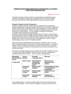
Transcription of London Cancer Lymphoma Radiotherpay Guidelines
1 London CancerGuidelines for themanagement of LymphomaJune Guidelines are intended to direct the treatment of patients with Lymphoma with radiotherapy. They havebeen developed from Guidelines already in existence at Barts Health NHS Trust, University College LondonHospitals NHS Foundation Trust, Royal Free London NHS Foundation Trust, Princess Alexander Hospital, NorthMiddlesex Hospital and Barking, Havering and Redbridge University Hospitals NHS Trust. They should be read andused in conjunction with other Guidelines covering the investigation and management of Hodgkins and Non-Hodgkins Lymphoma . They also do not remove the need to follow the Local Rules and Work Instructions that havebeen developed at individual radiotherapy System (Ann Arbor Staging Classification for Lymphoma )Stage IInvolvement of a single lymph node region (I).
2 Single extra nodal site (IE)Stage IIInvolvement of 2 or more lymph node regions on the same side of the diaphragm (II)Localised involvement of an extra lymphatic organ or site and its regional lymph node(s) with orwithout involvement of other lymph node regions on the same side of the diaphragm (IIE)Stage IIII nvolvement of lymph node regions on both sides of the diaphragm (III), which may beaccompanied by localised involvement of extra lymphatic site (IIIE) or spleen (IIIS) or both (IIIES)Stage IVDiffuse involvement of one or more extra lymphatic organs with or without associated nodalinvolvement or distant nodal involvement. Any liver / bone marrow diseaseXBulk disease>10cmA=absence of B symptomsB=presence of either unexplained fever>38 C; drenching night sweats; weight loss>10% in the 6months prior to ClassificationWHO Classification (Full extent is beyond the scope of this document).
3 Non-Hodgkins Lymphoma (NHL) Low Grade / Indolent ( Follicular, MALT) High Grade ( Diffuse Large B-Cell Lymphoma ; Primary Mediastinal; NK/T Cell Lymphoma )Hodgkins Lymphoma (HD) Classical HD: Nodular Sclerosing; Mixed Cellularity; Lymphocyte Deplete; Lymphocyte Rich Lymphocyte Predominant HD (LPHD)Early stage HDFavourableoClinical Stage I/II and no risk factorsUnfavourableoClinical Stage I/II with one or more of the following risk factors: Large mediastinal mass (>10cm) Extranodal involvement Elevated ESR (>30mm/h for B stage; >50mm/h for A stage) 3 or more lymph node regions involved B stage HD2 Favourable Clinical Stage III/IV with 0-3 adverse risk factors (listed below)Unfavourable Clinical stage III or IV with four or more adverse risk factorsoAlbumin level of < level of < of 45 years.
4 OStage IV blood cell (WBC) count of 15,000/mm3. oAbsolute lymphocytic count of <600/mm3 or a lymphocyte count that was <8% of the total WBC and INTENTR adical intentoFollowing short course chemotherapyoAs single modality therapy localized stage I Lymphocyte Predominant HD;oConsolidation following chemotherapyoChemotherapy refractory diseasePalliative Radiotherapy to start 4 weeks from time of decision to treat. Following chemotherapy recommendedstarts at maximum within 3 months of completing chemotherapy ( patient factors) but ideally to start4 weeks following completion. Lymphoma falls into Category 2 Dose schedulesClassical Hodgkin Lymphoma Favourable Early Stage after 2 cycles ABVD20Gy / 10 fractions over 2 weeks Unfavourable Early Stage after 4 cycles ABVD30Gy / 15-17 fractions Unfavourable Early Stage after BEACOPPDose may be reduced to 20Gy / 10 fractions 35-40Gy / 20 fractions may be considered in certain instances chemotherapy refractory Early stage, sole therapy30-35Gy / 15-20 fractions (dependent on bulk and site)Non-Hodgkin Lymphoma High grade Lymphoma30Gy / 15-17 fractions NK/ T cell Lymphoma requires higher doses of at least 50Gy in 2Gy/fraction Primary CNS Lymphoma - Post chemotherapy 35-40Gy in with boosting of residualvolume to total of 45-50Gy Low grade Lymphoma ( Follicular Lymphoma )
5 24-30 Gy / 12-15 fractions3 Examples of Palliative Schedules: 20-30Gy / 5-10 fractions 12Gy / 4 fractions 8Gy / single fraction 4Gy / 2 INVESTIGATIONS AND INFORMATION REQUIRED PRIOR TO DECISION TOTREAT FOR EBRTThe following investigations should have been performed with results available before planning commences: Clinical history Baseline clinical examination and Performance Status Histology FBC, U+E s, biochemical profile, LDH Bone marrow evaluation Results of staging investigations: CT and or FDG PET-CT. In some instances MRI may be of value CNS Lymphoma ; disease in head and neck region For patients receiving radiotherapy after chemotherapy, PET-CT / CT scans before and after treatment areused to determine the involved sites and residual disease. PET-CT pre and post chemotherapy is advisedfor ISRT (Involved Site Radiotherapy).
6 FOR PATIENTSP atients should be given an appropriate patient information leaflet about their treatment and have access to alymphoma nurse clinical nurse specialist or other specialist patients must have given written informed consent before radiotherapy planning commences. Consent is to betaken by a practitioner who is familiar with Lymphoma radiotherapy planning and IELSG 32 Randomised Phase2 trial of primary chemotherapy with high dose Methotrexate and high doseCytarabine with or without Thiopeta and with or without rituximab followed by brain radiotherapy vsHigh dose chemotherapy supported by Autologous stem cell transplant for immune-competent patientswith newly diagnosed primary CNS Lymphoma UK Haplo UK multi-centre phase 2 trial of haplo-identical stem cell transplantation in patients withhaematological malignancies RIC UCBTT ransplantation of umbilical cord blood from unrelated donors in patients with haematological diseasesusing a reduced intensity conditioning TREATMENT / IMMOBILISATION Patients to be planned and treated
7 In the supine position. Chin up position for neck and SCF sites. For head sites clinician to indicate appropriate neck position. Appropriate immobilization for the site being treated is required. In head and neck regions this shouldinclude a customized immobilization ACQUISITION Patients are 3D-planned using data from a CT planning scan. Contiguous slices with slice thickness of no more than 3mm taken through the region of interest. contrast is recommended to improve identification of nodal chains unless there are specific contra-indications. With common treatment planning systems, dosimetric calculations should not be influenced,except in sites such as mediastinum and para-aortic region where blood, volume is relatively large. Preand post contrast planning CT scans are then required.
8 It is recommended that each centre carry out adosimetric analysis of the effects of contrast on the treatment planning calculations for individualanatomical DELINEATION AND NOMENCLATUREL ymph node region atlases for CT planning have been published for major regions in the head and neck; trunk andpelvis and these should be referred to when outlining nodal Field Radiotherapy (IFRT) has been the standard with equivalence to wide field radiotherapy when usedin combination with Site Radiotherapy (ISRT) has been utilized in recent paediatric Hodgkin Lymphoma protocols and in therecent 18-30 trial as a step to further reduce the radiation volume treated and hence probability of late from large datasets is awaited from the current clinical et al, (2013) recommended the adoption of ISRT for patients receiving combined modality treatment aslong as appropriate pre-chemotherapy imaging is available.
9 In this instance, FDG PET-CT would be advisable. Ifimaging is not available or radiotherapy is being used as sole therapy, IFRT should be used instead. Use of ISRT remains at clinician discretion with the patient fully Gross tumour volumeCTV Clinical Target volumeIFRT CTV definition Involved field CTV (IF-CTV) will include the anatomical nodal region affected by Lymphoma defined by theclinician as that which should be treated by radiotherapy. IF-CTV will be outlined to include the involved nodal region, the margins of any tumour mass (primary orresidual) in all dimensions, & the contigual nodal regions. For patients who have had prior chemotherapy, the post chemotherapy volume is used in all directionsexcept cranio-caudal direction where the pre-chemotherapy volume is used There may be instances where it will be desirable to modify the IF CTV to limit toxicity.
10 This will beperformed under the clinician s discretion taking into account site of nodal regions are described with summary as follows:Neckipsilateral neck (mastoid suprasternal notch) including supraclavicular fossa (SCF)Mediastinumlower neck (from top of thyroid cartilage), bilateral SCF to5cm below lower extentof diseaseMediastinum + hilumas for mediastinum but includes bilateral hilar nodes; Inferior border (INF) tobottom of T10 SCFincludes ipsilateral neck. If mediastinum involved pre-chemo extend INF as permediastinum node regionAxillaincludes ipsilateral lower neck, SCF and infra-clavicular fossa (ICF) (top of thyroidcartilage to axillary fold)Inguinalipsilateral femoral, inguinal and external iliac node region; from bifurcation ofcommon iliacs to Sartorius muscle (at approx.)