Transcription of Lung‐RADS® Version 1
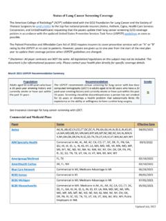
1 Lung RADS Version Assessment Categories Release date: 2019 Category Descriptor Lung-RADS Score Findings Management Risk of Malignancy Est. Population Prevalence Incomplete 0 Prior chest CT examination(s) being located for comparison Additional lung cancer screening CT images and/or comparison to prior chest CT examinations is needed n/a 1% Part or all of lungs cannot be evaluated Negative No nodules and definitely benign nodules 1 No lung nodules Continue annual screening with LDCT in 12 months < 1% 90% Nodule(s) with specific calcifications: complete, central, popcorn, concentric rings and fat containing nodules Benign Appearance or Behavior Nodules with a very low likelihood of becoming a clinically active cancer due to size or lack of growth 2 Perifissural nodule(s) (See Footnote 11) < 10 mm (524 mm3) Solid nodule(s): < 6 mm (< 113 mm3) new < 4 mm (< 34 mm3) Part solid nodule(s): < 6 mm total diameter (< 113 mm3) on baseline screening Non solid nodule(s) (GGN).
2 <30 mm (<14137 mm3) OR 30 mm ( 14137 mm3) and unchanged or slowly growing Category 3 or 4 nodules unchanged for 3 months Probably Benign Probably benign finding(s) short term follow up suggested; includes nodules with a low likelihood of becoming a clinically active cancer 3 Solid nodule(s): 6 to < 8 mm ( 113 to < 268 mm3) at baseline OR new 4 mm to < 6 mm (34 to < 113 mm3) 6 month LDCT 1 2% 5% Part solid nodule(s) 6 mm total diameter ( 113 mm3) with solid component < 6 mm (< 113 mm3) OR new < 6 mm total diameter (< 113 mm3) Non solid nodule(s) (GGN) 30 mm ( 14137 mm3) on baseline CT or new Suspicious Findings for which additional diagnostic testing is recommended 4A Solid nodule(s).
3 8 to < 15 mm ( 268 to < 1767 mm3) at baseline OR growing < 8 mm (< 268 mm3) OR new 6 to < 8 mm (113 to < 268 mm3) 3 month LDCT; PET/CT may be used when there is a 8 mm ( 268 mm3) solid component 5 15% 2% Part solid nodule(s): 6 mm ( 113 mm3) with solid component 6 mm to < 8 mm ( 113 to < 268 mm3) OR with a new or growing < 4 mm (< 34 mm3) solid component Endobronchial nodule Very Suspicious Findings for which additional diagnostic testing and/or tissue sampling is recommended 4B Solid nodule(s) 15 mm ( 1767 mm3) OR new or growing, and 8 mm ( 268 mm3) Chest CT with or without contrast, PET/CT and/or tissue sampling depending on the *probability of malignancy and comorbidities.
4 PET/CT may be used when there is a 8 mm ( 268 mm3) solid component. For new large nodules that develop on an annual repeat screening CT, a 1 month LDCT may be recommended to address potentially infectious or inflammatory conditions > 15% 2% Part solid nodule(s) with: a solid component 8 mm ( 268 mm3) OR a new or growing 4 mm ( 34 mm3) solid component 4X Category 3 or 4 nodules with additional features or imaging findings that increases the suspicion of malignancy Other Clinically Significant or Potentially Clinically Significant Findings (non lung cancer) S Modifier may add on to category 0 4 coding As appropriate to the specific finding n/a 10% IMPORTANT NOTES FOR USE: 1) Negative screen: does not mean that an individual does not have lung cancer 2) Size.
5 To calculate nodule mean diameter, measure both the long and short axis to one decimal point, and report mean nodule diameter to one decimal point 3) Size Thresholds: apply to nodules at first detection, and that grow and reach a higher size category 4) Growth: an increase in size of > mm (> 2 mm3) 5) Exam Category: each exam should be coded 0 4 based on the nodule(s) with the highest degree of suspicion 6) Exam Modifiers: S modifier may be added to the 0 4 category 7) Lung Cancer Diagnosis: Once a patient is diagnosed with lung cancer, further management (including additional imaging such as PET/CT) may be performed for purposes of lung cancer staging; this is no longer screening 8) Practice audit definitions: a negative screen is defined as categories 1 and 2; a positive screen is defined as categories 3 and 4 9) Category 4B Management: this is predicated on the probability of malignancy based on patient evaluation, patient preference and risk of malignancy.
6 Radiologists are encouraged to use the McWilliams et al assessment tool when making recommendations 10) Category 4X: nodules with additional imaging findings that increase the suspicion of lung cancer, such as spiculation, GGN that doubles in size in 1 year, enlarged lymph nodes etc 11) Solid nodules with smooth margins, an oval, lentiform or triangular shape, and maximum diameter less than 10 mm or 524 mm3 (perifissural nodules) should be classified as category 2 12) Category 3 and 4A nodules that are unchanged on interval CT should be coded as category 2, and individuals returned to screening in 12 months 13) LDCT.
7 Low dose chest CT *Additional resources available at - *Link to Lung-RADS calculator.