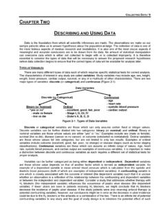
Transcription of MANAGEMENT OF BLUNT SPLENIC INJURY
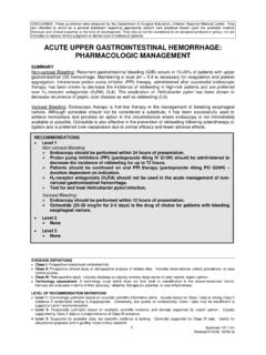
1 DISCLAIMER: These guidelines were prepared by the Department of Surgical Education, Orlando Regional Medical Center. They are intended to serve as a general statement regarding appropriate patient care practices based upon the available medical literature and clinical expertise at the time of development. They should not be considered to be accepted protocol or policy, nor are intended to replace clinical judgment or dictate care of individual patients. MANAGEMENT OF BLUNT SPLENIC INJURY . SUMMARY. SPLENIC INJURY can be initially managed with observation, angiographic embolization, or surgery depending upon the hemodynamic status of the patient, grade of SPLENIC INJURY , and presence of other injuries and medical comorbidities.
2 Evolving practice beginning in the pediatric population and expanding into adults, has allowed non-operative observation to become more prevalent for hemodynamically stable patients. Improvements in CT sensitivity and specificity have made vascular extravasation easier to diagnose, and interventional radiology has become an integral part of the MANAGEMENT of SPLENIC injuries, in some institutions replacing emergency operation as the treatment of choice. RECOMMENDATIONS. Level 1. In the hemodynamically stable patient, with suspected SPLENIC INJURY , IV contrast-enhanced CT. remains the gold standard diagnostic examination due to its speed, widespread availability, diagnostic accuracy, and relatively noninvasive nature.
3 In the hemodynamically unstable trauma patient with suspected SPLENIC INJURY , a positive FAST. scan or peritoneal signs requires emergent abdominal exploration to determine the source of intraperitoneal hemorrhage. Level 2. Non-operative MANAGEMENT in hemodynamically stable patients with BLUNT SPLENIC INJURY is considered standard of care. Angioembolization should be performed on all hemodynamically stable patients with contrast blush on CT scan of the abdomen. Routine angiography and embolization should be performed on hemodynamically stable patients with Grade IV-V SPLENIC INJURY . Level 3. Angiography may be used as an investigative tool to identify vascular abnormalities in patients who have sustained SPLENIC INJURY that have a continued drop in hemoglobin who remain hemodynamically stable, or pose a risk for delayed hemorrhage INTRODUCTION.
4 With regard to BLUNT trauma, the spleen is considered the most commonly injured intra-abdominal organ, accounting for up to 45% of all visceral injuries. Prior to the advent of CT scanning, physical examination and diagnostic procedures such as diagnostic peritoneal lavage (DPL) were the methods to guide surgical decision making. Minor SPLENIC INJURY was frequently missed, while major INJURY in the setting of hemodynamic instability and physical findings prompted emergent laparotomy. In early reports dated to the 1900s, immediate splenectomy was the standard of care for any degree of SPLENIC INJURY , a course of action that may have been influenced by previous assertions that the spleen is physiologically unnecessary.
5 Many studies, including one of King and Shumacker in 1952, demonstrated EVIDENCE DEFINITIONS. Class I: Prospective randomized controlled trial. Class II: Prospective clinical study or retrospective analysis of reliable data. Includes observational, cohort, prevalence, or case control studies. Class III: Retrospective study. Includes database or registry reviews, large series of case reports, expert opinion. Technology assessment: A technology study which does not lend itself to classification in the above-mentioned format. Devices are evaluated in terms of their accuracy, reliability, therapeutic potential, or cost effectiveness. LEVEL OF RECOMMENDATION DEFINITIONS.
6 Level 1: Convincingly justifiable based on available scientific information alone. Usually based on Class I data or strong Class II. evidence if randomized testing is inappropriate. Conversely, low quality or contradictory Class I data may be insufficient to support a Level I recommendation. Level 2: Reasonably justifiable based on available scientific evidence and strongly supported by expert opinion. Usually supported by Class II data or a preponderance of Class III evidence. Level 3: Supported by available data, but scientific evidence is lacking. Generally supported by Class III data. Useful for educational purposes and in guiding future clinical research.
7 1 Approved 09/30/2015. the importance of the spleen in immunity and the increased risk of infection among splenectomized patients (1). There were also misconceptions stemming from reports in the early part of the last century that non-operative MANAGEMENT (NOM) resulted in both a 90 100% mortality rate and a significant risk of delayed SPLENIC rupture. However, reports in the literature as early as 1939 described evidence of healed SPLENIC injuries at autopsy. Despite such reports, and various pioneering surgical attempts at SPLENIC salvage, splenectomy remained the standard of care through the early 1970s. This led in recent decades to a more conservative approach in the MANAGEMENT of SPLENIC injuries.
8 NOM of SPLENIC trauma in children was reported in 1968 by Upadhyaya, followed by other authors in the 1980s that have demonstrated good results with this method (2). Hemodynamic instability historically has been the primary reason for operative MANAGEMENT of SPLENIC trauma. The definition of hemodynamic instability varies, with many defining this as either a systolic blood pressure less than 90 mm Hg, need for ongoing resuscitation, or patients felt to have other associated injuries requiring operative MANAGEMENT . In the 1990s, CT scans became the gold standard for the diagnosis of solid organ injuries; this modality permitted identification of concomitant injuries, grading of SPLENIC injuries and broad quantification and imaging based comparisons of degrees of hemoperitoneum (3).
9 Further, the introduction of angioembolization increased the options available for SPLENIC salvage. To standardize the reporting of SPLENIC injuries, in 1994 the Organ INJURY Scaling Committee of the American Association for the Surgery of Trauma (AAST) developed a grading system based on the anatomic disruption of the spleen, as shown on CT scans or during laparotomy. The grading system is as follows. Grade I. o Hematoma: subcapsular, <10 percent of surface area o Laceration: capsular tear <1 cm in depth into the parenchyma Grade II. o Hematoma: subcapsular, 10 to 50 percent of surface area o Laceration: capsular tear, 1 to 3 cm in depth, but not involving a trabecular vessel Grade III.
10 O Hematoma: subcapsular, >50 percent of surface area OR expanding, ruptured subcapsular or parenchymal hematoma OR intraparenchymal hematoma >5 cm or expanding o Laceration: >3 cm in depth or involving a trabecular vessel Grade IV. o Laceration involving segmental or hilar vessels with major devascularization ( , >25 percent of spleen). Grade V. o Hematoma: shattered spleen o Laceration: hilar vascular INJURY which devascularizes spleen Technologic improvements have increased the accuracy of CT in identifying major SPLENIC injuries. Active SPLENIC hemorrhage is usually seen on current contrast-enhanced CT scans as an irregular or linear area of contrast extravasation.