Transcription of Management of Burns - WHO | World Health Organization
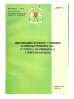
1 Management of Burns The Burns patient has the same priorities as all other trauma patients. Assess: - Airway - Breathing: beware of inhalation and rapid airway compromise - circulation : fluid replacement - Disability: compartment syndrome - Exposure: percentage area of burn. Essential Management points: - Stop the burning - ABCDE. - Determine the percentage area of burn (Rule of 9's). - Good IV access and early fluid replacement. The severity of the burn is determined by: - Burned surface area - Depth of burn - Other considerations. Morbidity and mortality rises with increasing burned surface area. It also rises with increasing age so that even small Burns may be fatal in elderly people. Continued next page WHO/EHT/CPR 2004 reformatted. 2007 WHO Surgical Care at the District Hospital 2003 1. Burn Management iinn A. Adduullttss The Rule of 9's is commonly used to estimate the burned surface area in adults.
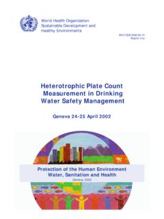
2 The body is divided into anatomical regions that represent 9% (or multiples of 9%) of the total body surface (Figure 7). The outstretched palm and fingers approximates to 1% of the body surface area. If the burned area is small, assess how many times your hand covers the area. Morbidity and mortality rises with increasing burned surface area. It also rises with increasing age so that even small Burns may be fatal in elderly people. Continued next page WHO/EHT/CPR 2004 reformatted. 2007 WHO Surgical Care at the District Hospital 2003 2. Burn Management iinn C. Chhiillddrreenn The Rule of 9's' method is too imprecise for estimating the burned surface area in children because the infant or young child's head and lower extremities represent different proportions of surface area than in an adult (see Figure 8). Burns greater than 15% in an adult, greater than 10% in a child, or any burn occurring in the very young or elderly are serious.
3 Continued next page WHO/EHT/CPR 2004 reformatted. 2007 WHO Surgical Care at the District Hospital 2003 3. Burn Management (continued). Depth of burn It is important to estimate the depth of the burn to assess its severity and to plan future wound care. Burns can be divided into three types, as shown below. Depth of Characteristics Cause burn First degree Erythema Sunburn burn Pain Absence of blisters Second degree Red or mottled Contact with hot (Partial Flash Burns liquids thickness). Dark and leathery Fire Third degree Dry Electricity or lightning (Full Thickness) Prolonged exposure to hot liquids/ objects It is common to find all three types within the same burn wound and the depth may change with time, especially if infection occurs. Any full thickness burn is considered serious. Serious burn requiring hospitalization - Greater than 15% Burns in an adult - Greater than 10% Burns in a child - Any burn in the very young, the elderly or the infirm - Any full thickness burn - Burns of special regions: face, hands, feet, perineum - Circumferential Burns - Inhalation injury - Associated trauma or significant pre-burn illness: diabetes Continued next page WHO/EHT/CPR 2004 reformatted.
4 2007 WHO Surgical Care at the District Hospital 2003 4. Burn Management (continued). Wound care First aid If the patient arrives at the Health facility without first aid having been given, drench the burn thoroughly with cool water to prevent further damage and remove all burned clothing. If the burn area is limited, immerse the site in cold water for 30 minutes to reduce pain and oedema and to minimize tissue damage. If the area of the burn is large, after it has been doused with cool water, apply clean wraps about the burned area (or the whole patient) to prevent systemic heat loss and hypothermia. Hypothermia is a particular risk in young children. First 6 hours following injury are critical; transport the patient with severe Burns to a hospital as soon as possible. Initial treatment Initially, Burns are sterile. Focus the treatment on speedy healing and prevention of infection.
5 In all cases, administer tetanus prophylaxis. Except in very small Burns , debride all bullae. Excise adherent necrotic (dead). tissue initially and debride all necrotic tissue over the first several days. After debridement, gently cleanse the burn with ( g/litre). chlorhexidine solution, (1 g/litre) cetrimide solution, or another mild water- based antiseptic. Do not use alcohol-based solutions. Gentle scrubbing will remove the loose necrotic tissue. Apply a thin layer of antibiotic cream (silver sulfadiazine). Dress the burn with petroleum gauze and dry gauze thick enough to prevent seepage to the outer layers. Continued next page WHO/EHT/CPR 2004 reformatted. 2007 WHO Surgical Care at the District Hospital 2003 5. Burn Management (continued). Daily treatment Change the dressing daily (twice daily if possible) or as often as necessary to prevent seepage through the dressing.
6 On each dressing change, remove any loose tissue. Inspect the wounds for discoloration or haemorrhage, which indicate developing infection. Fever is not a useful sign as it may persist until the burn wound is closed. Cellulitis in the surrounding tissue is a better indicator of infection. Give systemic antibiotics in cases of haemolytic streptococcal wound infection or septicaemia. Pseudomonas aeruginosa infection often results in septicaemia and death. Treat with systemic aminoglycosides. Administer topical antibiotic chemotherapy daily. Silver nitrate ( aqueous). is the cheapest, is applied with occlusive dressings but does not penetrate eschar. It depletes electrolytes and stains the local environment. Use silver sulfadiazine (1% miscible ointment) with a single layer dressing. It has limited eschar penetration and may cause neutropenia. Mafenide acetate (11% in a miscible ointment) is used without dressings.
7 It penetrates eschar but causes acidosis. Alternating these agents is an appropriate strategy. Treat burned hands with special care to preserve function. Cover the hands with silver sulfadiazine and place them in loose polythene gloves or bags secured at the wrist with a crepe bandage;. Elevate the hands for the first 48 hours, and then start hand exercises;. At least once a day, remove the gloves, bathe the hands, inspect the burn and then reapply silver sulfadiazine and the gloves;. If skin grafting is necessary, consider treatment by a specialist after healthy granulation tissue appears. Continued next page WHO/EHT/CPR 2004 reformatted. 2007 WHO Surgical Care at the District Hospital 2003 6. Burn Management (continued). Healing phase The depth of the burn and the surface involved influence the duration of the healing phase. Without infection, superficial Burns heal rapidly.
8 Apply split thickness skin grafts to full-thickness Burns after wound excision or the appearance of healthy granulation tissue. Plan to provide long term care to the patient. Burn scars undergo maturation, at first being red, raised and uncomfortable. They frequently become hypertrophic and form keloids. They flatten, soften and fade with time, but the process is unpredictable and can take up to two years. In children - The scars cannot expand to keep pace with the growth of the child and may lead to contractures. - Arrange for early surgical release of contractures before they interfere with growth. Burn scars on the face lead to cosmetic deformity, ectropion and contractures about the lips. Ectropion can lead to exposure keratitis and blindness and lip deformity restricts eating and mouth care. Consider specialized care for these patients as skin grafting is often not sufficient to correct facial deformity.
9 Nutrition Patient's energy and protein requirements will be extremely high due to the catabolism of trauma, heat loss , infection and demands of tissue regeneration. If necessary, feed the patient through a nasogastric tube to ensure an adequate energy intake (up to 6000 kcal a day). Anaemia and malnutrition prevent burn wound healing and result in failure of skin grafts. Eggs and peanut oil and locally available supplements are good. WHO/EHT/CPR 2004 reformatted. 2007 WHO Surgical Care at the District Hospital 2003 7.