Transcription of Management of distal clavicle fractures - Acta Orthopaedica
1 Management of type II distal clavicle fractures hasalways been a challenge. Non-operative treatment hasa high risk of complications and should be consideredonly for elderly and frail patients. For younger andactive patients there is a wide variety of operativeoptions, each with advantages and to our unit s experience the first choicecould be hook plate fixation, with very good andreproducible results. Another option could beKirschner-wire fixation with or without tension bandwiring ; however, because of potential wire complica-tions or difficulties in rehabilitation, the methodshould be reserved for reliable patients and used witha meticulous technique.
2 Keywords: distal clavicle fractures ; Management of fractures of the distal clavi-cle has been a matter of debate in literature. Neer in1968 suggested a new classification and proposedgeneral treatment guidelines(14). Type I and type IIIfractures are generally treated non-operatively. Fortype II fractures , although surgical Management isthe treatment of choice, some authors suggest conservative treatment owing to the high rate ofcomplications in particular in 1968 classified distal clavicle fracturesaccording to their location in relation to the coraco-clavicular ligaments(14).
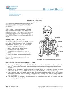
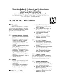
3 Type I fractures are stablefractures located lateral to the coracoclavicularligament complex. Type II fractures are complexunstable fracture -dislocations which leave the distalend of the clavicle and the acromioclavicular (AC)joint untouched, separating the clavicle from theunderlying coracoclavicular ligament complexthrough a vertical or oblique fracture line (fig 1).Type III fractures are intra-articular fractures intothe AC joint causing late posttraumatic arthritis andpain(14).
4 Rockwood, in 1982, subclassified type II fractures in type IIa and IIb fractures . In type IIafractures, the fracture line is located medial to thecoracoclavicular ligaments and both trapezoid andconoid ligaments remain connected to the distalfragment. In type IIb fractures , the fracture line islocated between the coracoclavicular ligaments :the trapezoid ligament remains intact whereas theconoid ligament is ruptured(19,22).No benefits or funds were received in support of this studyActa Orthop dica Belgica, Vol.
5 76 - 2 - 2010 Acta Orthop. Belg., 2010, 76, 145-149 Management of distal clavicle fracturesIlias BISBINAS, Petros MIKALEF, Ioannis GIGIS, Theodoros BESLIKAS, Nikolaos PANOU, Ioannis CHRISTOFORIDISFrom Aristotle University of Thessaloniki, GreeceREVIEW ARTICLE Ilias Bisbinas, MD, Orthopaedic Consultant. Petros Mikalef, MD, Orthopaedic Consultant. Ioannis Gigis, MD, Lecturer in Orthopaedics. Theodoros Beslikas, MD, Assistant Professor. Nikolaos Panou, MD, Orthopaedic Consultant. Ioannis Christoforidis, MD, Professor and Department, Aristotle University ofThessaloniki, G.
6 Gennimatas General Hospital, EthnikisAminis, Thessaloniki, : Ioannis GIGIS, Profiti Ilia 13, Pylea,55535, Thessaloniki, Greece. E-mail : 2010, Acta Orthop dica in 1998 suggested a further classifica-tion combining the above mentioned principles ; ithas not been widely accepted(20).TreatmentTreatment of type I and type III fractures doesnot appear to be debated : most authors suggestconservative treatment with a period of immobiliza-tion in a sling followed by physiotherapy(3).
7 Apotential late complication of intra-articular type IIIfractures is post-traumatic AC joint arthritis ; whenthis is symptomatic, excision of the lateral end ofthe clavicle is suggested(19).There is a wide variety of treatment options forNeer type II fractures , all of them based on the par-ticular deformity and lack of stability. Bone frag-ments are displaced due to forces that act in oppo-site directions. The weight of the upper extremity,pull of the latissimus dorsi, pectoralis major andminor muscles, as well as rotation of the scapuladisplace the distal clavicular fragment downwardsand medially, while upon moving the upper extrem-ity, the distal fragment rotates and tilts even trapezius muscle pulls the proximal fragmentupwards and backward.
8 As a result of the displace-ment of proximal and distal bone fragments, malu-nion is commonly seen after non-operative treat-ment(10,21,23). Most authors consider that the highrisk of complications observed after conservativemanagement makes operative treatment the mostprudent treatment consists of open reductionand internal fixation (ORIF) of the fracture . Severalmethods of fixation have been suggested, butnone of them has been considered the goldstandard (19,22,24).
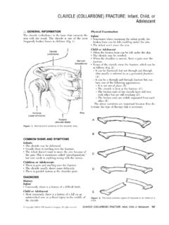
9 Transacromial fixation usingKirschner wires, cerclage wiring of the fragments,tension band wiring, plate fixation, osteosynthesisusing a hook plate (fig 2), stabilization of the medi-al fragment with either a coracoclavicular screw orcoracoclavicular slings have been mentioned in lit-erature(5,19,22,24). The fixation hardware is usuallyremoved 8 weeks postoperatively as soon as radio-logical consolidation of the fracture is observed. Allthose operative methods have their own advantagesand transacromial fixation, Kirschner wires orKnowles pins are introduced through the lateralside of the acromion after open reduction of thefracture(4,5,6).
10 Disadvantage of the method is thatthe stabilization of the fracture is poor, and earlymobilization of the arm is therefore not suggest-ed(9). The reported infection and non-union ratesare high, up to 23%(6). Injury to the AC jointmay be followed by arthritis, with a rate of 10%.The incidence of pin migration is reportedlyhigh(1,5,13). Lyons and Rockwood in 1990reviewed all reports about pin migration in the ACjoint surgery in literature(13).