Transcription of Mandibular first molar with four canals in the mesial root
1 CLINICAL. Mandibular first molar with four canals in the mesial root Hamed Karkehabadi1, Ricardo Machado2 and Lucas da Fonseca Roberti Garcia3. Abstract A 35-year-old female patient with intermittent pain in the lower right jaw was referred to the dental practice. The right Mandibular first molar did not respond to thermal testing and the patient reported moderate pain to percussion and palpation in the region of this tooth. Radiographic findings confirmed the presence of periradicular disease and previous root canal treatment. Initially, only four treated canals were found (two mesial and two distal) Two extra canals were found in the mesial root. The treatment was performed and the success confirmed after 12 months.
2 This case report reiterates the complexity of Mandibular first molar variation and is intended to reinforce the clinicians'. need to be aware of the variable morphology of root canals in this tooth. Introduction The goal of endodontic treatment is to clean and shape the root canal system and obturate it in all its dimensions (Almeida et al, 2015). Before starting treatment, aberrant root or root canal morphology should be investigated, detected and adequately treated (Lea et al, 2014). Failure to recognise any unusual canal configuration could eventually lead to an unsuccessful treatment outcome (Almeida et al, 2015; Lea et al, 2014;. Ghoddusi et al, 2007). Thus, the proper knowledge of the root and root canal morphology, along with any related anatomical variations, is essential to achieve a successful treatment (Almeida et al, 2015; Lea et al, 2014; Ghoddusi et al, 2007).
3 Mandibular first molars are the teeth that most require endodontic treatment, as they are the first posterior teeth to erupt in permanent dentition (Ghoddusi et al, 2007). Although Mandibular first molars usually have two canals in the mesial root, named as mesiobuccal and mesiolingual canals , the presence of a middle mesial canal in the developmental groove has been widely reported in 1 % (Vertucci, 1964), (Martinez-Berna and Badanelli, 1983), % (Fabra-Campos, 1985), (Fabra- Campos, 1989), 12% (Pomeranz et al, 1981), (Akbarzadeh et al, 2017), 15%. (Goel et al, 1991) and (Versiani et al, 2016) of the cases. In addition, 1. Hamed Karkehabadi Mandibular first molars may have one or two canals in the distal root (Ghoddusi et al, Department of endodontics, dental school, Hamadan University of 2007).
4 Medical Sciences, Hamadan, Iran As reported in several studies, the prevalence of a middle mesial canal in Mandibular first molars may significantly range among researches. Clinical studies and case reports 2. Ricardo Machado that demonstrated the presence of negotiable middle mesial canals present different Clinical practice limited to endodontics in Navegantes, data from studies which used extracted teeth model (Vertucci, 1984; Martinez-Berna Santa Catarina, Brazil and Badanelli, 1983; Fabra-Campos, 1985; Fabra-Campos, 1989; Pomeranz et al, 1981, Akbarzadeh et al, 2017; Goel et al, 1991; Versiani et al, 2016). 3. Lucas da Fonseca Roberti Garcia To the best of the authors' knowledge, just one case has ever been reported in the Department of dentistry - endodontics division, Federal scientific literature of a Mandibular first molar with four canals in the mesial root (Reeh, University of Santa Catarina, 1998) In this particular case, the author reported a successful endodontic retreatment Florian6polis, Santa Catarina, Brazil of a Mandibular first molar with seven canals , where the other three canals were located 42 INTERNATIONAL DENTISTRY AFRICAN EDITION VOL.
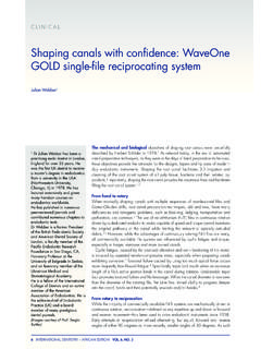
5 8, NO. 6. CLINICAL. Case Report 1. 1a 1b 1c 1d 1e 1f 1g 1h Figure 1: Initial radiograph (a); Radiographic working length confirmation (b); Radiograph of main cones (c); Final radiographs (d- f); Radiographs of 12-month follow-up (g and h). in the distal root. Initially, the clinician failed to detect the three canals were treated endodontically. Such factors complexity of the root canal system of this tooth, and only demonstrate the importance of knowing the root canal system VOL. 8, NO. 6 INTERNATIONAL DENTISTRY AFRICAN EDITION 43. KARKEHABADI ET AL. morphology and its anatomical variations (Reeh, 1998). 20 file. Irrigation was performed with ml of Furthermore, according to the author of the case report, sodium hypochlorite solution (Chloraxid) at each change of looking for additional canals was crucial for a successful file, using a 30-gauge, open-ended needle (Navitip.)
6 Outcome (Reeh, 1998). Ultradent, South Jordan, UT, USA) placed slightly short of the Therefore, the aim of this article is to report a clinical case of binding point. After a final rinse with MTAD (Dentsply Tulsa, a Mandibular first molar with four canals in the mesial root, and USA), the root canals were dried with sterile absorbent paper two canals in the distal root. All canals were properly prepared points (Dentsply Sirona, Ballaigues, Switzerland) and a and filled, and the success of the treatment was confirmed after radiograph was taken of the main cones to confirm if they 12 months by clinical and radiographic findings. reach the working length (Figure lc). Afterwards, the canals were filled with gutta percha (Dentsply Sirona, Ballaigues, Case report Switzerland) and AH Plus sealer (Dentsply Sirona, Ballaigues, A 35-year-old female patient, with a history of intermittent Switzerland), using the lateral condensation technique.
7 The prior to being seen, was referred to the dental practice. pulp chamber was properly cleaned to remove the excess Medical history was non-contributory. The right Mandibular of gutta percha and sealer, and was temporarily restored first molar did not respond to thermal testing (heated gutta with a provisional material (Figures 1d -1f). percha and dry ice), and the patient reported moderate pain Clinical and radiographic findings, as no painful to percussion and palpation in the region of this tooth. symptomatology and regression of the periapical lesion, Furthermore, the tooth had a total crown and there was no confirmed the success of the proposed therapy. Continued significant probing depth.
8 Radiographic findings confirmed follow-up over 12 months has shown a positive outcome from the presence of periradicular disease and previous root canal endodontic perspective (Figures Ig and Ih). treatment (Figure la). Correlation of clinical and radiographic findings led to a diagnosis of a chronic periradicular abscess Discussion due to previous unsuccessful endodontic treatment. Therefore, The prevention and treatment of apical periodontitis are the a nonsurgical root canal retreatment was recommended. main objectives of endodontic therapy (Siqueira and R as, After administrating infiltration anaesthesia (Lidocaine 2% 2008). An adequate biomechanical preparation and filling 1 epinephrine), the crown was removed, and of the root canal system, associated to the placement of a rubber dam isolation of the operative area was performed.
9 Proper coronal sealing, are crucial and should be taken to An endodontic access cavity was prepared by using achieve this goal (Gillen et al, 201l). Furthermore, all these 1016HL and Endo Z burs (Dentsply Sirona), and four steps performed during the treatment require a good previously treated root canals were found. Removal of root understanding of root canal anatomy to attain a successful canal filling was performed using a Neoniti Al size 20 file outcome (Reeh, 1998). (Neolix, Ch tres-la-For t, France). After careful analysis of The purpose of this article was to present the case report the floor of the pulp chamber, two additional root canals of a Mandibular first molar clearly showing the presence of were found in the mesial root, totalling four canals in this four canals in the mesial root, and two canals in the distal root.
10 Initially, extra canals were explored with a size 10 root. The patient was referred to the dental practice, because k-file (Mani, Inc; Tochigi, Japan) under copious irrigation with of the persistent symptoms after the first endodontic treatment, sodium hypochlorite solution (Chloraxid, Cerkamed, at which time, only four root canals were located and treated Poland). Next, the working length of each root canal was (two in the mesial root and two in the distal root). established 1mm up to the apical foramen, using an The clinician should keep in mind that the internal electronic apex locator (Root ZX, Morita, Tokyo,Japan). A morphology of the teeth does not always follow the known radiograph was then taken to confirm the measurements standards (Almeida et al, 2015; Lea et al, 2014).