Transcription of MDCT PWV - j-ca.org
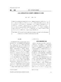
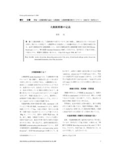
1 Online publication May 27, 2010. 49 2 . MDCT PWV .. CT. MDCT .. MDCT . MDCT . PWV .. J Jpn Coll Angiol, 2010, 50: 163 167 . Key words: coronary artery disease, multi-detector row computed tomography, coronary calcification, pulse wave velocity 97 . PPV NPV . 87 . 99 1 .. arterial stiffness .. >50 .. 91 96 PPV 76 NPV. 99 2 NPV 100 .. MetS .. MDCT .. CT. MDCT . Fuster . ACS .. PWV . 3 .. MDCT . ACS . MDCT .. Fig. 1 .. 64 MDCT CT . Raff 94 Inoue ACS . 2009 3 27 . THE JOURNAL of JAPANESE COLLEGE of ANGIOLOGY Vol. 50, 2010 163. MDCT PWV . Figure 1 Volume rendering image and multi-planner reconstruction image were shown in A and B, respectively. Severe stenosis (A, B) and plaque with positive remodeling (B). was identified in the proximal left anterior descending coronary artery.
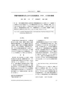
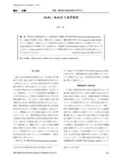
2 CT 50 HU ACS . CT ACS .. 25 15 HU vs. 71 16 HU .. Fig. 2 . 4 . MDCT Mo- toyama ACS 30 HU . spotty . 5 . MDCT .. MDCT . Figure 2 Comparison of the CT density of plaques between . Fig. 3 . 2 . patients with acute coronary syndrome (ACS) and stable angi- MDCT na (SA). In the culprit lesions, the mean CT density of the plaque was 25 15 HU (from -12 to 48 HU) in patients with .. ACS and 71 16 HU (from 46 to 101 HU) in patients with SA. MDCT There was a statistically significant difference between the 2. groups (p < ).. MDCT . 3 . 0 12 10 .. 13 445 > 445 1,000 . 12 . 2 6 .. 3 .. Fig. 4 Budoff . 2 . MDCT . 164 Vol. 50, 2010. 2 . Figure 3. A: A case of non-calcified plaque (CAC score = 0) with significant coronary stenosis in LAD observed by MDCT. and CAG. B: A case of moderate calcification (CAC score =.)
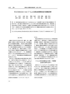
3 245). Coronary stenosis was not diagnosed by MDCT. because of calcification and no significant coronary stenosis was confirmed by CAG. C: A case of severe calcification (CAC score = 3,809). Coronary stenosis was not diagnosed by MDCT because of severe calcification. Significant coronary stenosis in LAD was confirmed by CAG. CAC, coronary artery calcification; MDCT, multi-detector row computed tomography; CAG, coronary angiography;. LAD, left anterior descending coronary artery.. PWV .. PWV .. Moens-Korteweg .. 7 . vascular damage . Figure 4 Coronary artery calcification (CAC) score according to the number of significant stenosed vessels (VD) evaluated by invasive coronary angiography. The CAC score in 3VD was signifi- . cantly higher than in the other groups (0, 1VD and 2VD).
4 PWV 8, 9 PWV * p < vs. 0, 1 and 2. Vol. 50, 2010 165. MDCT PWV . Table 1 Contributor of CAC score groups as assessed by logistic regression analysis Variable p value Upstroke time (mean) < Sex Age PWV (mean) HDL-C Hypertension %MAP (mean) CCB Systolic blood pressure ARB Figure 5 Correlations between brachial-ankle pulse wave velocity (baPWV) and length of abdominal aortic calcification Uric acid (AAC). The length of AAC was measured on X-ray films, and Diabetes mellitus this has been reported to correlate significantly with cardiovas- PWV, pulse wave velocity; MAP, mean arterial pressure; CCB, cular morbidity and mortality. baPWV was significantly corre- calcium channel blocker; ABI, ankle/brachial pressure index; PWV, lated with AAC length. pulse wave velocity; ARB, angiotensin II receptor blocker.
5 Based on reference 12.. Fig. 5 . 10 . IMT . 11 .. PWV 1 Leschka S, Alkadhi H, Plass A et al: Accuracy of MSCT. coronary angiography with 64-slice technology: rst expe- MDCT rience. Eur Heart J, 2005, 26: 1482 1487. PWV 2 Mitsutake R, Niimura H, Miura S et al: Clinical signi cance PWV of the coronary calci cation score by multidetector row com- PWV puted tomography for the evaluation of coronary stenosis in Japanese patients. Circ J, 2006, 70: 1122 1127.. up- 3 Fuster V, Badimon L, Badimon JJ et al: The pathogenesis of . stroke time, UT . coronary artery disease and the acute coronary syndromes.. Table 1 . 12 . N Engl J Med, 1992, 326: 310 318. PWV 4 Inoue F, Sato Y, Matsumoto N et al: Evaluation of plaque texture by means of multislice computed tomography in pa- MDCT tients with acute coronary syndrome and stable angina.
6 Circ PWV J, 2004, 68: 840 844. UT 5 Motoyama S, Kondo T, Sarai M et al: Multislice computed tomographic characteristics of coronary lesions in acute coronary syndromes. J Am Coll Cardiol, 2007, 50: 319 326. 6 Budoff MJ, Shaw LJ, Liu ST et al: Long-term prognosis associated with coronary calcification: observations from MDCT . a registry of 25,253 patients. J Am Coll Cardiol, 2007, 49: MDCT 1860 1870. 7 Yamashina A, Tomiyama H, Takeda K et al: Validity, repro- PWV ducibility, and clinical signi cance of noninvasive brachial- MDCT ankle pulse wave velocity measurement. Hypertens Res, 2002, 25: 359 364. 166 Vol. 50, 2010. 2 . 8 Lehmann ED, Riley WA, Clarkson P et al: Non-invasive abdominal aortic calcification. Hypertens Res, 2003, 26: assessment of cardiovascular disease in diabetes mellitus.
7 163 167. Lancet, 1997, 350 Suppl 1: SI14 SI19. 11 Matsumoto M, Inoue K, Moriki A: Associations of brachial- 9 Cruickshank K, Riste L, Anderson SG et al: Aortic pulse-wave ankle pulse wave velocity and carotid atherosclerotic lesions velocity and its relationship to mortality in diabetes and with silent cerebral lesions. Hypertens Res, 2007, 30: 767 773. glucose intolerance: an integrated index of vascular function? 12 Mitsutake R, Miura S, Saku K: Association between coronary Circulation, 2002, 106: 2085 2090. artery calci cation score as assessed by multi-detector row com- 10 Nakamura U, Iwase M, Nohara S et al: Usefulness of brachial- puted tomography and upstroke time of pulse wave. Inernt ankle pulse wave velocity measurement: correlation with Med, 2007, 46: 1833 1836.
8 Association between Coronary Stenosis or Calcification Score on MDCT and PWV. Ryoko Mitsutake, Shin-ichiro Miura, and Keijiro Saku Department of Cardiology, Fukuoka University School of Medicine, Fukuoka, Japan Key words: coronary artery disease, multi-detector row computed tomography, coronary calcification, pulse wave velocity Multi-detector row computed tomography (MDCT) has become more widely available in many general hospitals, as it enables the accurate non-invasive assessment of coronary artery stenosis, plaque imaging and calcium scoring. However, it is very difficult to detect coronary stenosis on MDCT in a lesion with advanced calcification. We analyzed the association between coronary stenosis or calcification score on MDCT and pulse wave velocity, and found additional information for diagnosis of coronary artery disease.
9 (J Jpn Coll Angiol, 2010, 50: 163 167). Online publication May 27, 2010. Vol. 50, 2010 167.