Transcription of Medial Patellofemoral Ligament Repair and Reconstruction ...
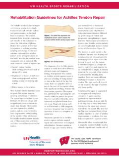
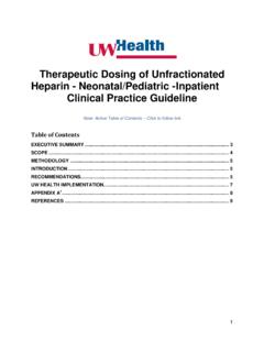
1 Figure 1. Radiograph of the Patellofemoral joint with the knee in slight flexion. The lateral aspect of the trochlear groove is normally about 1 cm higher than the Medial . Medial Patellofemoral Ligament Repair and Reconstruction Proper stabilization of the patella is also affected by the soft tissue structures (ligaments and muscles) surrounding the knee. The Medial Patellofemoral Ligament (MPFL) is a continuation of the deep retinaculum and vastus medialis oblique (VMO) muscle fibers (inner portion of the quadriceps muscle) on the inside of the knee. These structures provide a significant force (near 60% total) against lateral displacement of the patella, as their force is directed inward or ,4 The MPFL is the primary restraint to lateral displacement of the patella during the first 20 to 30 degrees of knee This Ligament is a passive stabilizer and extends from the upper inner side of the patella to the Medial aspect of the femur.
2 The patellomeniscal Ligament and retinaculum also contribute more than 20% of the restraining force. The knee consists of four bones that form three joints. The femur is the large bone in the thigh and attaches by ligaments and a capsule to the tibia, the large bone in the lower leg commonly referred to as the shin bone. Next to the tibia is the fibula, which runs parallel to the tibia on the outside of the leg. The patella, commonly called the kneecap, is embedded in the quadriceps and patellar tendon which articulates with the front of the femur, which forms the Patellofemoral joint. The patella acts as a pulley to increase the amount of force that the quadriceps muscle can The patella sits in a groove on the end of the femur called the trochlear groove.
3 This groove varies in depth from person to person. When the knee bends, the patella travels down the groove and as the knee straightens the patella moves up the groove. As the patella travels up and down in the trochlear groove, the patella should maintain congruent boney alignment, which is often referred to as normal patellar tracking. There are several structures that work together to keep the patella aligned and stabilized in the femoral groove to prevent the patella from excessive lateral movement (movement towards the outside of the leg). The lateral aspect of the trochlear groove is normally about 1 centimeter higher than the Medial (inside of the leg) aspect of the trochlear groove, which helps keep the patella in the trochlear groove by providing a buttress on the lateral side (Figure 1).
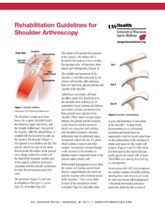
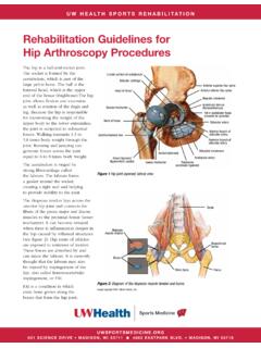
4 2 This provides the main resistance to lateral patellar translation (which is the most common direction of displacement), especially beyond 20 degrees of knee People who have a shallow trochlea are more susceptible to patellar instability. 2 These ligaments can be injured and torn with an initial acute traumatic patellar dislocation (kneecap quickly going out of place). The most common mechanism for a patellar dislocation is a forceful inward rotation of the body on a planted foot. This radiograph is of a 12-year-old boy in the emergency room after such an injury (Figure 2) Often the patella will go back in to place (or relocate to the groove) as the knee is gently straightened. In this case the patient was unable to straighten his knee in the emergency room and his patella was still dislocated laterally.
5 Note on the radiograph that there is no overlap of the femur and patella. In studying 26 patients who had an acute patellar dislocation at a mean age of 18 years, Nomura et al. reported evidence of MPFL damage in 96% (26/27) of the knees examined during open surgical exploration). In an acute patellar dislocation, when a tear of the MPFL is identified, surgical Repair (fixing the original Ligament ) of the MPFL may be a good treatment option. In the young athletic population, recurrence rates for patients treated conservatively are high with some studies reporting 40%. In recurrent or chronic patellar dislocations, it may be necessary to perform Reconstruction of the MPFL. Reconstruction differs from Repair in that graft tissue (such as a hamstring tendon) is used to replace or reinforce the MPFL.
6 The MPFL Reconstruction may also be combined with other patellar stabilization procedures. A quality post-operative rehabilitation program is essential to having a successful outcome from a MPFL procedure. The goals of rehabilitation will initially focus on protection for healing, mobility and range of motion. After this early phase, strengthening and neuromuscular control is emphasized throughout the entire leg and core. In the final stages of rehabilitation, the focus will be on dynamic lower extremity control during sport specific movements, such as change of direction and rotational movements. The UW Health Sports Medicine rehabilitation guidelines below are presented in a criterion-based progression.
7 General time frames are given for reference to the average, but individual patients will progress at different rates depending on their age, associated injuries, pre-injury health status, rehabilitation compliance and injury severity. Modifications in the specific time frames, restrictions and precautions may also be made to protect healing tissues based on the specific surgical Repair / Reconstruction procedure performed. 3 Pha se I (surgery to 6 weeks a fter surgery) Rehabilitation appointments Rehabilitation appointments to begin 2-3 days after surgery and to continue 1-2 times per week Rehabilitation goals Protect surgical knee Restore normal knee range of motion Full passive knee extension should be achieved within first two weeks, thought these patients do not often struggle with extension Goal of a 90 degrees knee flexion should be achieved by 6 weeks Normalize gait Eliminate effusion (swelling) Restore leg control and quadriceps activation Precautions Weight bearing as tolerated on crutches with post-operative brace locked in extension.
8 Begin ambulation with 2 crutches, then progress to 1 crutch and then no crutches once patient shows sufficient quad control and gait mechanics are normalized Avoid rotational movements through knee and lower extremity Suggested therapeutic exercise Gait drills with emphasis on symmetrical loading, heel strike at initial contact, appropriate quad activation during stance (avoid hyperflexed or hyperextended knee during mid-stance), and adequate push-off Range of motion (ROM): (pearl: provide manual lateral patellar stabilization when first initiating range of motion) Knee extension with foot propped on bolster Heel slides Knee flexion wall slides Passive knee flexion over edge of plinth Medial Patellar mobilizations Prone knee flexion Strengthening: Note: it is recommended that all quadriceps strengthening be performed in conjunction with neuromuscular electrical stimulation (NMES).
9 Please see Appendix for recommendations on NMES units for patients, parameters for use, and treatment recommendations for NMES during quadriceps strengthening. Quadriceps, hamstring, and gluteal sets 4 Four-way leg lifts in standing or lying down positions for hip strengthening Bridging Ankle isotonics with resistance band Weight shifting drills Heel raises balance drills beginning with double leg and progressing to single leg Trunk stability work Supine core and transverse abdominus activation with upper and/or lower extremity movement Anti-rotation press variations in stable lower extremity positions Suitcase carry with gait and/or marching drills Cardiovascular exercise Upper body circuit training or use of an upper body ergometer Progression criteria Non-painful knee flexion AROM to 90 degrees Full weight bearing with normalized gait
10 Mechanics without the use of assistive device Single leg balance for 15 seconds with good control 5 Phase II (begin after meeting Phase I criteria, usually 6 weeks after surgery) Rehabilitation appointments Rehabilitation appointments are 1-2 times per week Rehabilitation goals Maintain/fully restore normal knee range of motion Maintain protection of post-surgical knee Increase functional activity Good limb control and no pain with functional movements First Progressive Testing session should occur at 12 weeks after surgery: LSI >90% on single leg press and Y- balance Quadriceps strength deficit of <30% on Biodex strength test Precautions Use of lateral buttress knee sleeve if directed by physician or physical therapist Avoid over-stressing fixation of graft continued caution with rotational movement For patients with patellar or trochlear chondroplasty or cartilage defect.