Transcription of Membrane Potential
1 7. Membrane Potential The Resting Membrane Potential Results From the tances. These electrical signals-receptor potentials, Separationof ChargesAcross the Cell Membrane synaptic potentials, and action potentials-are all pro- The Resting Membrane Potential Is Determined by Resting duced by temporary changes in the current flow into Ion Channels and out of the cell that drive the electrical Potential RestingChannelsin Glial Cells Are Selectivefor acrossthe cell Membrane away from its resting value. PotassiumOnly This current flow is controlled by ion channels in RestingChannelsin Nerve Cells Are Selectivefor Several the cell Membrane . We can distinguish two types of ion Ion Species channels-resting and gated-by their distinctive roles PassiveFlux of Sodium and PotassiumIs Balancedby in neuronal signaling. Resting channels normally are Active Pumping of the Ions open and are not influenced significantly by extrinsic Chloride Ions May Be PassivelyDistributed factors, such as the Potential acrossthe are primarily important in maintaining the resting The Balanceof Ion Fluxes That Gives Rise to the Resting Membrane Potential Is Abolished During the Action Membrane Potential , the electrical Potential across the Potential Membrane in the absenceof signaling.
2 Most gated chan- The Contributions of Different Ions to the Resting nels, in contrast, are closed when the Membrane is at Membrane Potential Can Be Quantified by the Goldman rest. Their probability of opening is regulated by the Equation three factors we considered in the last chapter: changes The Functional properties of the Neuron Can Be Represented in Membrane Potential , ligand binding, or Membrane in an electrical Equivalent Circuit stretch. EachIon ChannelActs as a Conductor and Battery in In this and succeeding chapters we consider how Parallel transient electrical signals are generated in the neuron. We begin by discussing how resting ion channelsestab- An Equivalent Circuit Model of the MembraneIncludes Batteries,Conductors,a Capacitor,and a Current lish and maintain the resting Potential . We also briefly Generator describe the mechanism by which the resting Potential An Overall View can be perturbed, giving rise to transient electrical sig- nals such as the action Potential .
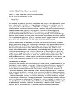
3 In Chapter 8 we shall consider how the passive properties of neurons -their resistive and capacitive characteristics-contribute to I NFORMATION IS CARRIEDWITHIN and between neurons by electrical and chemical signals. Transient electri- local signaling within the neuron. In Chapter 9 we shall examine how voltage-gated Na +, K+, and Ca2+ chan- nels generate the action Potential , the electrical signal conveyed along the axon. Synaptic and receptor poten- cal signals are particularly important for carrying tials are considered in Chapters 10-13 in the context of time-sensitive information rapidly and over long dis- synaptic signaling between neurons . 126 Part II / Cell and Molecular Biology of the Neuron The Resting Membrane Potential Results .. (j(j(I(j(j( .. ( . 1. From the Separation of Charges Across Equal the Cell Membrane Every neuron has a separation of chargesacrossits cell +,- leG). ( I. Extr8cellular Membrane consisting of a thin cloud of positive and 8<iCia<t$ ~ '".)))))))
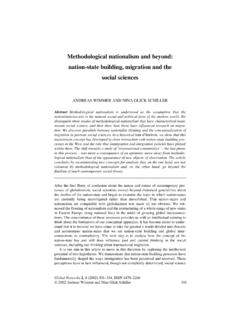
4 ". side negative ions spread over the inner and outer surfaces -':1, ,." -:.. of the cell Membrane (Figure 7-1). At rest a nerve cell Cytoplasmic has an excessof positive charges on the outside of the Membrane and an excessof negative chargeson the in- Q. side side. This separation of charge is maintained because the lipid bilayer of the Membrane blocks the diffusion of ions, as explained in Chapter 6. The charge separation .Q \$. 8( . G.. gives rise to a difference of electrical Potential , or volt- age, acrossthe Membrane called the membranepotential. Figure 7-1 The Membrane Potential results from a separa- The Membrane Potential (Vm) is defined as tion of positive and negative charges across the cell mem- brane. The excess of positive charges (red circles) outside the Vm = Vin - Vout, membraneand negativecharges (blue circles) inside the mem- brane of a nerve cell at rest representsa small fraction of the total number of ions inside and outside cell.)
5 Where VIn is the Potential on the inside of the cell and Voutthe Potential on the outside. The Membrane Potential of a cell at rest is called the We begin examining the Membrane Potential by an- , by convention, the po- alyzing how the passive flux of individual ion species tential outside the cell is defined as zero, the resting po- through resting channelsgeneratesthe resting Potential . )tential (Vr) is equal to VIn' Its usual range in neurons is We shall then be able to understand how the selective -60 mY to -70 mY. All electrical signaling involves gating of different types of ion channels generatesthe brief changesfrom the resting Membrane Potential due i,\ctionpotential, as well as the receptor and synaptic po- to alterations in the flow of electrical current acrossthe tentials. cell Membrane resulting from the opening and closing of ion channels. The electric current that flows into and out of the The Resting Membrane Potential Is cell is carried by ions, both positively charged (cations) Determined by Resting Ion Channels and negatively charged (anions).
6 The direction of cur- rent flow is conventionally defined as the direction of No single ion speciesis distributed equally on the two net movement of , in an ionic solu- $ides of a nerve cell Membrane . Of the four most abun- tion cations move in the direction of the electric current, (tant ions found on either side of the cell Membrane , anions in the opposite direction. Whenever there is a net Na+ and CI- are more concentrated outside the cell, flow of cations or anions into or out of the cell, the and K+ and organic anions (A -) are more concentrated charge separation across the resting Membrane is dis- inside. The organic anions are primarily amino .acids turbed, altering the polarization of the Membrane . A re- and proteins. Table 7-1 shows the distribution of these duction of charge separation, leading to a less negative ions inside and outside one particularly well-studied Membrane Potential , is called increase nerve cell process, the giant axon of the squid, whose in charge separation, leading to a more negative mem- blood has a salt concentration similar to sea water.)
7 Al- brane Potential , is called in though the absolute values of the ionic concentrations Membrane Potential that do not lead to the opening of for vertebrate nerve cells are two- to threefold lower gated ion channels, are called electrotonicpotentialsand t1hanthosefor the squid giant axon, the concentration are said to be passive responsivesof the Membrane . Hy- gradients(the ratio of the external ion concentration to perpolarizing responsesare almost always passive , as internal ion concentration) are about the same. are small depolarizations. However, when depolariza- The unequal distribution of ions raises several im- tion approachesa critical level, called the threshold,the portant questions. How do ionic gradients contribute to cell responds actively with the opening of voltage-gated the resting Membrane Potential ? How are they main- ion channels, which at threshold produces an all-or- tained?What prevents the ionic gradients from dissipat- none actionpotential(Box 7-1).
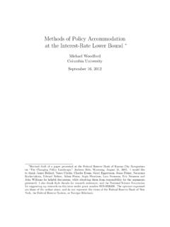
8 Ing by diffusion of ions across the Membrane through Chapter 7/ MembranePotential 127. [embrane Potential g the electrical Potential across inside of the Membrane becomesmore positive while the out- l in the late tech- side of the Membrane becomes more negative. This progres- ~ of both the resting and the sive decreasein the normal separation of charge is called dep0- ! of glass micropipettes filled larization. ion that serve as electrodes. :ed on either side of the cell the back ends of the pipettes to an oscilloscope, which dis- nbrane Potential in volts. Be- microelectrode is very small l cell with relatively little dam- :~ . ~~ I--t Inwerd TIme - 5 Oms Figure 7-2C Depolarization. Small depolarizing current pulses evoke purely electro- tonic ( passive ) potentials in the cell-the size of the change in Potential is proportional to the size of the current pulses. How- NeMIcell ever, sufficiently large depolarizing current triggers the open- ing of voltage-gated ion channels.]
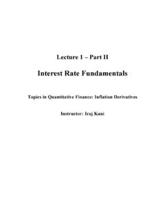
9 The opening of these chan- ecording setup. nels leads to the action Potential , which differs from electrotonic potentials not only in the way in which it is gener- Itside the cell no electrical p0- ated but also in magnitude and duration. ut as soon as one microelec- Reversing the direction of current flow-making the in- ~oscilloscope shows a steady tracellular electrode negative with respect to the extracellular otential. In most nerve cells at electrode-makes the Membrane Potential more negative. round -65 mY. This increasein chargeseparation is called hyperpolilrization. ,Iectrode lOng Potential I':l me- oscope display. n be experimentally changed cted to a second pair of elec- )ne extracellular. When the Figure 7-2D Hyperpolarization. c>sitivewith respect to the ex- re current from the generator The responsesof the cell to hyperpolarization are usually Ie neuron from the intracellu- purely electrotonic-as the size of the current pulse increases, , to the extracellular electrode the hyperpolarization increasesproportionately.
10 Hyperpolar- Membrane . As a result, the ization does not trigger an active responsein the cell. 128 Part n / Cell and Molecular Biology of the Neuron Table 7-1 Distributionof the Major IonsAcrossa NeuronalMembraneat Rest:the GiantAxon of the Squid Concentration Concentration in Equilibrium Species of in cytoplasm extracellular fluid potential1. ion (mM) (mM) (mV). -75. +55. -60. the passive (resting) channels? These questions are The separation of charge resulting from the diffusion of interrelated, and we shall answer them by considering K+ gives rise to an electrical Potential difference: posi- two examples of Membrane permeability: the resting tive outside, negative inside. The more K+ continues to Membrane of glial cells, which is permeable to only one flow, the more charge will be separated and the greater speciesof ions, and the resting Membrane of nerve cells, will be the Potential difference. Since K+ is positively which is permeable to three.