Transcription of MRI of the IAC’s: Value of a Dedicated Scan with …
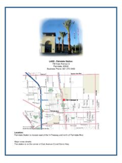
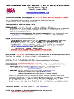
1 Accredited by the American aic College of Radiology ADVANCED IMAGING CENTER. PHYSICIAN NEWS. April 18, 2012. MRI of the IAC's: Value of a Dedicated scan with Contrast Fig. 1a Fig 1b Fig 2a Fig. 2b CLINICAL PRESENTATION: This patient presented with a history of right ear pain and hearing loss. The ABR test had come out abnormal. An MRI of the Brain and Dedicated MRI of the Internal Auditory Canals (IACs) were requested. IMAGING FINDINGS: The brain MRI was completely normal. Even on the post contrast images of the brain, no obvious abnormality could be detected. Fig. 1a-b: These are thin section axial T2 and T1 weighted images of the IAC's. They also show no obvious abnormality. Fig. 2a-b: These are thin section axial and coronal T1 weighted images of the IAC's AFTER Gadolinium injection. They reveal a subtle 2-mm enhancing lesion within the right IAC (arrows).. DISCUSSION: MRI of the brain is the modality of choice for almost all brain conditions.
2 In certain cases, a higher- resolution Dedicated scan of another area in the brain becomes indicated. Examples include Dedicated MRI of the IAC's with /without (w/wo) contrast, dynamic contrast MRI of the pituitary glands, high-resolution MRI of the hippocampus in seizure disorders, etc. The above example clearly demonstrates that if a Dedicated MRI of the IAC's ( with contrast) had not been performed, this abnormality would have been missed on the brain MRI w/wo contrast or even on a pre-contrast IAC scan . The 2-mm lesion in the right IAC could only be detected on the post contrast thin sections of the IAC. DIFFERENTIAL DIAGNOSIS: The DDX includes an early acoustic neuroma (schwanoma). A focal area of neuritis is less likely as neuritis tends to cover a longer length of the nerve. Since the lesion is very small (2 mm), it might be safe to follow this up with serial MRI's to rule out growth.
3 If the patient's symptoms are worsening, then surgical excision or radiation ablation (eg, with a Gamma knife) may be indicated. So if you have a patient with asymmetric hearing loss or dizziness and need to rule out a potential Acoustic Neuroma, then please order an MRI of the Brain (to rule out other brain pathologies) AND Internal Auditory Canals (IAC's) with and without contrast. Please do not hesitate to contact me should you have any questions. et [t {x |? `W. Ray H. Hashemi, , Diplomat American Board of Radiology ~ 43731 N. 15th St. West ~ Lancaster, CA 93534 ~ Tel: (661) 949-8111 ~ Fax: (661) 949-6600 ~ ~ 25842 Tournament Road ~ Valencia, CA 91355 ~ Tel: (661) 255-0060 ~ Fax: (661) 255-0024 ~ ~ 607 West Ave Q ~ Palmdale, CA 93550 ~ Tel: (661) 456-2020 ~ Fax: (661) 456-2021 ~ ~ 900 Heritage Dr., Bldg B ~ Ridgecrest, CA 93555 ~ Tel: (760) 446-1999 ~ Fax: (760) 446-1910]}