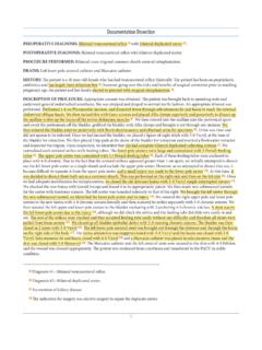
Transcription of MS Neck slide20 plain - AAPC
1 1 Documentation DissectionPREOPERATIVE DIAGNOSIS: Thoracic tumorPOSTOPERATIVE DIAGNOSIS: Secondary thoracic cancer from right breast : Right thorax exploration for excision of recurrent breast cancer : General FLUIDS: 3000 ml of BLOOD LOSS: 300 : Right cervical mass sent to Pathology, which is presumed recurrent right breast cancer : Jackson-Pratt in right : : : Ms. is a 72-year-old woman with breast cancer of the right breast who presented with right arm weakness and pain last year. At that time, she underwent a right brachial plexus exploration with resection of the metastasis. At that time, it was felt that we had no clean margins; however, since that time, she initially woke up neurologically intact.
2 Over the last few weeks to months, she has had a progressive pain in her right neck and right arm and first in her thumb as well as in her anterior lateral aspect of the right arm. She also was severely weak with only 1 to 2 out of 5 in her right deltoid and 2 to 3 in her right biceps. The MRI revealed large recurrence of tumor centered in the right brachial plexus that we felt had involved the upper trunk and was resulting in causing her weakness. Because of the severe pain and progressive deficits, risks and benefits of surgical exploration of the anterior thorax with re-resection were explained to the patient versus conservative management.
3 She elected to proceed with surgical resection. Risks and benefits of the procedure were explained to the patient and her family, and they elected to OF PROCEDURE: After informed consent was obtained, the patient was taken to the operating room and placed under general anesthetic. A standard surgical time-out was performed, and a dose of preoperative antibiotics was administered, at this time, with exposure below her right neck . Her existing Z-shaped incision was marked with plans to extend it in both directions. This region was prepped and draped in the usual sterile fashion and infiltrated with local anesthetic. There was one area of the thorax where tumor had obviously eroded to the skin as well as an area of wound dehiscence.
4 A decision was made to ellipse out both these regions and close them primarily at the conclusion of the case. This incision was opened with a #15 blade scalpel and carried down in subcutaneous tissues with Metzenbaum scissors. Immediately upon ellipsing out the tumor lesion as expected, there was a 5 x 6 cm tumor. A dissection was carried medially in an attempt to get around this aspect of the tumor and was taken up to the sternocleidomastoid muscle. The part of the SCM had to be resected to limit to remove this initial portion of tumor, which ultimately could be removed with what appeared to be clean margins. This tumor was sent off to Pathology as superficial tumor portion.
5 For the majority of the tumor, the dissection was carried down and the tumor was easily identified, Dissection was initially carried out in the superior direction, with quite a nice plane in the superior aspect of the tumor. Using Metzenbaum scissors and bipolar cautery, tissue plane was identified, and we were able to get around the tumor in a cephalad direction. This was repeated on the lateral margin and was taken all the way posterior down to the trapezius muscle. A nice, clean, fatty margin was identified and dissection was carried out on the lateral aspect of the tumor. On the inferior aspect of the tumor, it was carried down to the clavicle.
6 In a subclavicular fashion, the dissection was carried down until the tumor was encroaching on the subclavian vessels. Subclavian artery was identified and great care was taken to not disturb these vessels. Tumor was able to be peeled off with a nice margin in inferior direction as well. There appeared to be some abnormal tissue abutting the subclavian artery, but the risk of debriding this tumor was not worth with the damage of the artery. This erythematous area was left. Medially, dissection was carried around and this was where the margin was quite obscured. Slowly, it was taken around until we identified the phrenic nerve. The tumor was completely encasing the phrenic nerve and the decision was made to take nerve as we felt there would be no long-term #10 JP was placed in the cavity and tunneled through a separate stab exit site.
7 The dermal layer was reapproximated with 3-0 Vicryl in a buried interrupted fashion. A 3-0 nylon and horizontal mattress were used to close the skin. Sterile dressings were applied to the wound and the drain exit site was also secured down with a 3-0-nylon suture. Large fluffs and a compressive dressing were applied to the the conclusion of our portion of the case, all counts were correct x : To PACU in satisfactory HOL O G Y: confirmation of recurrent right breast cancer metastasis. What are the CPT and ICD-10-CM codes reported?


