Transcription of N-Glycan Analysis of Monoclonal Antibodies - IPT Online
1 Innovations in Pharmaceutical Technology Issue 4222 Laboratory TechnologyKeywordsN-glycan analysisProtein glycosylationMonoclonalantibodies (mAbs)Ultra high performance liquid chromatography (UHPLC)Fluorescence detectionBy Sonja Schneider and Edgar N gele at Agilent TechnologiesA Quaternary LC System, together with fluorescence detection, provides an optimal method for the sensitive and high-resolving Analysis of 2-AB derivatised glycans released from mAbs and other Analysis of Monoclonal AntibodiesProtein glycosylation is one of the most frequently observed post-translational modifications. Mammalian glycoproteins contain three major types of glycans: N-linked, O-linked and glycosylphosphatidylinositol (GPI) lipid anchors, which consist of one or more monosaccharide units.
2 A single glycosylation site can generate considerable heterogeneity regarding mass and charge of glycoproteins. These oligosaccharides are involved in many biological regulation and recognition processes, for example, protein sorting, immune and receptor recognition, inflammation, pathogenicity, metastasis, and other cellular processes. Therefore, certain glycosylation patterns can be associated with the diseased or healthy state of a patient (1). In addition, properties such as safety, efficacy and the serum half-life of therapeutic proteins can be affected by their glycosylation pattern. Recombinant Monoclonal antibody therapeutics (mAbs) represent the largest group of therapeutic proteins, representing a major new class of drug.
3 The efficacy of these therapeutics is highly dependent on the correct glycosylation patterns of the mAbs and, so far, all licensed therapeutic mAbs are immunoglobulin G (IgGs) (2). Human IgG has a single conserved N-linked glycosylation site located on the Fc region of each heavy chain at Asn-297 (3). This fact results in two sugar moieties per IgG, which are highly heterogeneous and contain up to 30 different glycan types (4). The combination of glycans at each of the two glycosylation sites on the Fc region leads to large numbers of different glycoforms in each batch of mAb glycan structure on this glycosylation site plays a critical role in complement activation and receptor affinity (5), which in turn affects the efficacy of the therapeutic mAbs.
4 Moreover, non-human glycans represent a safety issue due to induced immune responses. Therefore, Analysis of the glycan pattern is an important part of characterisation of therapeutic glycoproteins, especially mAbs. Figure 1a shows the general nomenclature used to describe sugar residues of different glycan structures on proteins. Figure 1b shows the predominant glycan structures present on the Asn-297 site in IgG. In general, N-glycans have a core structure, containing two `-D-N-acetylglucosamine (GlcNac) and three mannose (Man) units. IgG Fc N-glycans are predominantly biantennary complex-type structures, partially core fucosylated (for example, G0F). Different strategies for the Analysis of glycans have been described in the literature.
5 A large number of methods are based on protein-released and subsequently derivatised glycans due to the lack Images: Agilent Technologies, IncG0G0FG1G2 Figure 1: Glycan structure and isoforms: a) General nomenclature for glycans; b) Predominant glycan structures of IgGs. G = Galactose units, F = Fucose units. Modified after Arnold et al (3) in Pharmaceutical Technology Issue 42 Figure 2:Separation of mAb N-glycans with names, masses, monosaccharidecomposition and structure given for the mAb glycansare available in the N-Glycan Analysis application note (8)).Deglycosylation ProcedureDeglycosylation of the Monoclonal antibody and the glycoproteins was performed using PNGase F to release asparagine-linked oligosaccharides (N-glycans) from the glycoproteins.
6 PNGase cleaves asparagine-linked high mannose, as well as hybrid and complex oligosaccharides from the glycoproteins and leaves the glycans intact. The average glycosylation of proteins is 2-5 per cent with 1,000 Da as the average molecular weight. Therefore, to release approximately 20 g glycans, 400 g of glycoproteins was and conalbumin have only one glycosylation site, whereas the mAb contains two glycosylation sites and so the amount of PNGase F was adjusted to the amount of glycans. The proteins were deglycosylated for three hours at 37 C. The reaction was then stopped and the sample was vacuum dried for further processing. AB-Labelling for Fluorescence Detection and Sample Clean-upThe dried glycan samples were labelled with 2-AB using the GlycoProfi l 2-AB Labeling Kit according to the preparation protocol from Sigma-Aldrich for three hours at 65 C.
7 After the labelling procedure, the samples were purified using the GlycoProfi l Glycan Clean-up Cartridges from Sigma-Aldrich. After the HILIC clean-up procedure, the samples were vacuum dried and reconstituted in 15 L ultrapure water for liquid chromatography (LC) chromophores needed for optical detection methods, for example UV have presented a combination of enzymatic release of N-glycans using PNGase F with subsequent derivatisation with 2-aminobenzamide (2-AB) for fluorescence detection. 2-AB is a neutral and stable bonded label, often used in glycan Analysis (for example 6,7). The 2-AB-labelling is achieved by reductive amination, and results in a mass of the glycans of +119 Da. Due to protonation, the resulting mass shift is 120 Da (see Figures 2-4).
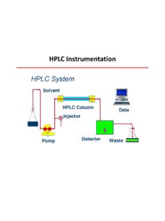
8 Due to the hydrophilic properties of glycans, subsequent purification using Hydrophilic Interaction chromatography Solid Phase Extraction (HILIC-SPE) is added for removal of excess label (6). Separation using HILIC with fluorescence detection is a robust method for glycan Analysis . Pauline Rudd and co-workers established a database ( ) based on HIILIC retention properties, a frequently used technology for the Analysis of protein glycosylation (6,7).To identify the monosaccharide composition of the glycans within a chromatographic peak, HILIC-LC can be coupled to electrospray Ionisation Quadrupole Time-of-Flight mass Spectrometry (ESI-QToF-MS) for mass and structure Glycan AnalysisHere, we demonstrate the Analysis of N-linked glycans with HILIC separation using the Agilent 1260 Infinity Bio-inert Quaternary LC System, together with the Agilent 1260 Infinity Fluorescence Detector.
9 (Full details of the components, equipment and software used along with chromatographic conditions LU86420 NameCalculated MassMonosaccharideStructuremass+ (1,6) (1,3) 0A 0A 0A (1,6) G1F(1,3) G1FG2F2120 0A 1A 2A in Pharmaceutical Technology Issue glycan pattern of the mAb standard showed a high amount of glycan G0, which was added as internal standard for system checkout. Figure 2 shows an overview of the detected glycan structures. The high intensity of the detected labelled glycans was achieved by setting the optimal wavelengths for glycan detection on the 1260 Infinity Fluorescence Detector, using 260 nm as the excitation wavelength and 430 nm as the emission wavelength. Usually, an excitation wavelength of 330 nm is preferred for the Analysis of 2-AB labelled glycans.)
10 We used the lower 260 nm, due to a higher intensity, resulting in better signal-to-noise ratios, as described by Melmer et al (7). Two additional glycoproteins (ovalbumin and conalbumin) were deglycosylated, and the glycans derivatised and then analysed using HILIC-UHPLC. Figure 3 shows the separation of ovalbumin glycans. Ovalbumin is N-glycosylated only at one site (Asn-292), but a complex glycosylation pattern can be associated with this site. Due to the complexity of the glycan pattern, the gradient had to be adjusted to achieve a higher resolution. Six glycans were identified with ESI-QTOF detection and mass correlation to the glycan library published by Harvey et al.(9). Over 35 peaks released from ovalbumin could be resolved in the glycan 4 shows the glycosylation pattern of conalbumin (synonym: ovotransferrin).