Transcription of Nerve Damage and Third Molar Removal - …
1 O R A L S U R G EO R A L. R Y S U R G E R Y. Nerve Damage and Third Molar Removal A. R. LOESCHER, K. G. SMITH AND P. P. ROBINSON. connective tissue and other structures in Abstract: The surgical Removal of lower Third molars endangers both the lingual and the neurovascular bundle. After injury, inferior alveolar nerves. Patients sustaining an injury to either of these nerves must be unless the Nerve is displaced into the managed correctly, and this requires a diagnosis of the injury type and regular monitoring of the recovery of sensation. Surgical intervention for a damaged inferior socket, the severed Nerve ends do not alveolar Nerve is not usually indicated but may be undertaken: if the Nerve is retract, but will remain in apposition. completely divided and the severed ends are misaligned; if a bony fragment has Regeneration within the canal will thus be compressed the mandibular canal; or if the patient suffers from persistent neuropathic unimpeded unless obstructed by pain.
2 In contrast, after injury to the lingual Nerve , if sensory testing demonstrates no displaced fragments of bone from the neural recovery within 3 4 months, exploration of the injury site and microsurgical roof of the canal. Good recovery after repair of the damaged Nerve is indicated. injury would therefore be expected. Many studies have reported the Dent update 2003; 30: 375 382 frequency of Nerve injury during the Clinical Relevance: It is imperative that patients sustaining a Nerve injury are Removal of Third molars (for review see managed correctly and this must include a diagnosis of the type of injury, monitoring Robinson 19971) and most indicate that recovery and the treatment of appropriate cases. inferior alveolar Nerve function is disturbed after 4 5% of procedures (range ). Most patients will regain normal sensation within a few weeks or months and less than 1% (range I mpacted mandibular Third Molar teeth are in close proximity to the lingual, inferior alveolar, mylohyoid and buccal speech and mastication and may adversely affect the patient's quality of life.)
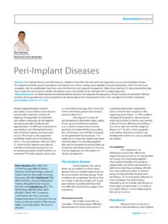
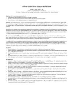
3 They also constitute one of the most 0 ) have a persistent sensory disturbance. nerves (Figure 1). During surgical frequent causes of complaints and The anatomical relationship between Removal , each of these nerves is at risk of litigation. It is therefore imperative that Damage , but the most troublesome patients sustaining Nerve injuries are complications result from inferior alveolar managed correctly, and this includes or lingual Nerve injuries. The majority of correct diagnosis of the type of injury, injuries result in transient sensory monitoring recovery, and the treatment of disturbance but, in some cases, appropriate cases. permanent paraesthesia (abnormal The characteristics of the inferior sensation), hypoaesthesia (reduced alveolar, lingual and buccal nerves, and sensation) or, even worse, some form of the likely incidence of Damage , will now dysaesthesia (unpleasant abnormal be discussed. Injury to the mylohyoid sensation) can occur (Table 1).
4 These Nerve is rare and is not considered further sensory disturbances can be in this brief update . troublesome causing problems with INFERIOR ALVEOLAR Nerve . Loescher, BDS, PhD, FDS RCS, MBChB, Smith, BDS, PhD, FDS RCS and The inferior alveolar Nerve is P. P. Robinson, BDS, PhD, FDS RCS, DSc, FMedSci, morphologically unusual in that it travels Department of Oral and Maxillofacial Surgery, a significant distance within bone in the School of Clinical Dentistry, Claremont Crescent, Figure 1. Cross-section of the mandible through mandible. In the mandibular canal it is the Third Molar showing the inferior alveolar, Sheffield S10 2TA. supported by the surrounding lingual, mylohyoid and long buccal nerves. Dental update September 2003 375. O R A L S U R G E R Y. In addition to the surgical technique, Anaesthesia Absence of all sensory modalities. other risk factors have been identified. Hypoaesthesia Diminished sensitivity to stimulation, excluding special senses.
5 Lingual Nerve Damage is particularly associated with deeply impacted teeth Paraesthesia An abnormal sensation, whether spontaneous or evoked. when the surgery is consequently Dysaesthesia Unpleasant abnormal sensation, whether spontaneous or evoked. difficult, particularly if distal bone Removal is ,10 The results of Hyperalgesia An increased response to a stimulus that is normally painful. studies comparing the incidence of Allodynia Pain due to a stimulus that does not normally provoke pain. lingual Nerve injury during surgery utilizing bone Removal with burs or Table 1. Pain terms adapted from the definitions of the International Association for the Study of Pain ( ). chisels are ,10 It is possible that the elevation of a lingual mucoperiosteal flap when chisels are utilized is of more the inferior alveolar Nerve and the roots trapped or constricted by scar tissue. importance than the method of bone of the Third Molar may be judged Regeneration of axons across a gap will Removal itself.
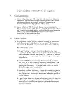
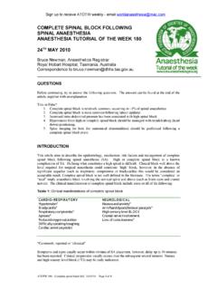
6 A recently published radiographically2,3,4,5 in an attempt to be less successful than if the Nerve ends prospective study undertaken by Renton predict the likelihood of Nerve Damage . remain in apposition. In addition, the and McGurk14 reported that factors Rood and Shehab (1990)5 identified five presence of a range of functionally reflecting the surgical skill ( lingual radiographic features that were related to distinct Nerve fibre types in this Nerve plate perforation) and the difficulty of the inferior alveolar Nerve injury and these ( mechanosensitive, thermosensitive, extraction were the strongest predictors are listed in Figure 2. The same study gustatory, vasomotor and secretomotor) of temporary and permanent lingual Nerve failed to show that narrowing of the may make successful regeneration of the injury. inferior alveolar canal' and the presence axons back to the correct receptor/. of a dark and bifid apex' were significant effector and location less likely (see predictors of inferior alveolar Nerve below).
7 BUCCAL Nerve . injury; features that had previously been There is a wide range in the reported This Nerve descends between the two considered Paradoxically, frequency of lingual Nerve injuries during parts of the lateral pterygoid muscle, sensory disturbance may occur following Third Molar Removal , with 22% of medial to the ramus of the mandible, and Removal of teeth that show none of these patients reporting sensory disturbances then passes laterally across the external features, and may not occur even when in the early post-operative period and 0 oblique ridge distal to the Third Molar , to there appears to be clear evidence of a 2% a permanent disturbance (for review supply the The sensory close relationship between the canal and see Robinson 19971). There are several distribution is variable but includes the the tooth roots. possible explanations for the wide range lower posterior buccal sulcus and A higher incidence of inferior alveolar in incidence.
8 First, the variation may Nerve injury has been reported with Third reflect differences in the time interval molars that are horizontally3,7 or between tooth Removal and the mesioangularly4 impacted and have assessment of the sensory impairment;. complete bone One study has early assessments will report many also demonstrated that increasing age is transient sensory changes that recover associated with a higher frequency of rapidly and completely, and which would inferior alveolar Nerve injury (14 24 year- be missed if assessment takes place after old patients ; 35 81 year-old a longer recovery period. Secondly, the patients ).8 incidence of Nerve injury may depend upon whether the sensory deficit was established objectively by the clinician or LINGUAL Nerve was based on a subjective patient The lingual Nerve is morphologically very assessment. Finally, it may reflect different from the inferior alveolar Nerve . differing surgical techniques; several Figure 2.
9 Five radiographic signs suggesting At the usual site of injury (adjacent to the studies have shown that the raising and juxtaposition of the mandibular canal to the Third lower Third Molar ) the Nerve is covered retraction of a lingual mucoperiosteal flap Molar roots, as described by Rood and Shehab with only a thin layer of soft tissue and is associated with an increased frequency Signs significantly related to Nerve injury: mucosa, rather than being in a bony of lingual Nerve ,10 Two recent A. Radiolucency across the roots of the Third canal. Consequently, if sectioned, the cut studies and a systematic review have Molar ; B. Deviation of the mandibular canal; C. Interruption of the white line of the canal. Signs Nerve ends retract apart and, if the concluded that raising and retracting a considered to be clinically important: D. adjacent soft tissue is also distorted, the lingual periosteal flap is not necessary11 Deflection of the Third Molar roots by the canal; E.
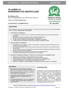
10 Nerve ends may become misaligned and and is best ,13 Narrowing of the Third Molar root. 376 Dental update September 2003. O R A L S U R G E R Y. classification is valuable as it emphasizes segmental demyelination, but these the importance of each structural changes are Recovery of component of the Nerve trunk (Figure sensation normally occurs within a few 3).21 Each of the five degrees of Nerve days of the surgical trauma, although injury, described by Sunderland, may be recovery may be slightly slower if created during the Removal of an segmental demyelination takes impacted Third Molar tooth: The cellular events that follow axonal l Compression injuries (Figure 3A) discontinuity (2nd to 5th degree injuries). may occur during the elevation of a have been studied extensively21,24 and are Third Molar with roots in close summarized in Figure 4. Wallerian proximity to the mandibular canal. degeneration occurs distal to the site of Minor compression of the Nerve , or the injury and usually extends centrally first degree injuries, will give rise to for a few This axonal a temporary conduction block, which degeneration consists of disintegration is referred to by Seddon19 as of both the axon and myelin, neurapraxia.