Transcription of Nostocoida limicola I - Activated Sludge,Process …
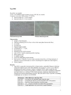
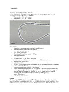
1 1 Nostocoida limicola I Resembles: N. limicola III: more robust filaments "Cand. M. parvicella": filaments do not stain grey-violet with Neisser Several Alphaproteobacteria: hybridise with probe ALF-968 Probes: not available, see remarks Frequency occurrence (200 samples; 175 WTPs): observed with a FI 1 in 1 sample observed with a FI 3 in 1 sample N. limicola I plus two M. parvicella filaments Characteristics bent/coiled, frequently tangled filaments; not motile; not branched; filament length > 200 m; cell diameter - m; sheath absent; attached growth absent; septa usually hardly visible; disc shaped or spherical cells; no sulphur storage; Gram positive; Neisser positive: filaments stain entirely grey-violet.
2 Remarks The Nostocoida limicola story is a little bit complicated and confusing. Three, Gram and Neisser positive, N. limicola morphotypes can be distinguished by applying phase-contrast microscopy [3]: N. limicola I: cell septa hardly visible; diameter m N. limicola II: cell septa visible; spherical to disc shaped cells; diameter: m 2 N. limicola III: cell septa visible; discoid to disc shaped cells; diameter: m. Note: The larger diameter range mentioned in reference 4 was based on outdated evidence and is therefore not valid any longer. As N. limicola II is not very often observed and, moreover, closely resembles N.
3 limicola III, it has been proposed to use the latter name for both morphotypes [4]. However, the name N. limicola II is still mentioned in published papers. Filamentous morphotypes resembling N. limicola filaments are frequently observed in WTPs treating industrial wastewater. It is now known that many of these morphotypes belong to the Alphaproteobacteria. Group specific (ALF-968) and several species specific probes are available and are needed to distinguish them from the N. limicola morphotypes. Several papers have been published in recent years in which it is stated by the authors that pure cultures of N. limicola resembling filaments were obtained [2, 7, 8, 11].
4 Subsequently, probes were developed based upon these pure cultures [9, 11,12]. 1. It has been assumed that probe NLIMI-91 should hybridise with N. limicola I [9, 12]. A fluorescent signal with this probe was obtained in 12 out of 126 Dynafilm samples. The size of the population giving a signal was usually small (1 on a scale ranging from 0 to 4). The probe positive filaments were, one sample excluded, very short (length < 50 m) and composed of almost spherical cells with a diameter of ca. m. Thus, the appearance of the probe NLIMI-91 positive, short filaments present in the Dynafilm samples was very similar to that in the original paper [9].
5 Considering that morphotype N. limicola I looks completely different (long curled and tangled filaments with a diameter of circa m), it is concluded that probe NLIMI-91 does not hybridise with N. limicola I. Filaments with the appropriate N. limicola I morphology, were observed in just one Dynafilm sample. These filaments did not hybridise with probe NLIMI-91. Hence, it is concluded that identification of morphotype N. limicola I through FISH is still not possible. A German research group reached a similar conclusion [10]. Probe NLIMI-91 can only be used for the identification of Trichococcus species, filamentous micro-organisms which have only a minor role in bulking of Activated sludge .
6 2. Probe NLIMII-175 was developed starting with sequences of pure cultures, described in reference 2 as "Candidatus N. limicola ", which were classified as members of the Actinobacteria. The pure cultures stained Gram and Neisser positive. This probe should allow the in situ identification of N. limicola II [9, 12]. Filaments hybridising with this probe were present in 9 Dynafilm samples and they were identified by phase-contrast microscopy as N. limicola III filaments. Thus, probe NLIMII-175 cannot be used for the in situ identification of morphotype N. limicola II, but targets N. limicola III. 3. Probe NLIMIII-301 should allow the identification of N.
7 limicola III through FISH [8, 9, 12]. A fluorescent signal with this probe was obtained in 13 samples. The filaments hybridising with probe NLIMIII-301 were rather short and composed of spherical cells with a diameter ranging from m to m. These filaments did not at all resemble the characteristic long, tangled N. limicola III filaments, composed of disc shaped cells, but instead those of a bacterium known as an Isosphaera sp. This conclusion is in agreement with the evidence presented in reference 10. 4. Ten Nostocoida limicola -like pure cultures, with an almost identical 16S rRNA sequence were isolated from domestic WTPs in Germany [11].
8 They were classified as members of the Chloroflexi (green non-sulphur bacteria). The pure cultures stained Gram positive and Neisser negative. Based upon their sequence, probe AHW-183 was developed and applied for the in situ identification of filaments in Activated sludge . Fluorescent signals were only obtained with domestic sludges. The filaments hybridising with AHW-183 indeed closely resemble those of morphotype N. limicola . They were mainly observed inside sludge flocs. Considering the Neisser negative staining results, it seems likely that this probe does not hybridise with N. limicola I or III. Unfortunately, probe AHW-183 was not applied during the Dynafilm research program.
9 3 In conclusion, when the Alphaproteobacteria are excluded, four N. limicola morphotypes can be observed in Activated sludge : N. limicola I: long, coiled and tangled filaments with a diameter of ca. m. The cell septa are hardly visible. Gram and Neisser positive. The phylogenetic position is not known and a probe is not yet available. N. limicola II: compared with N. limicola III, more spherical shaped cells and a somewhat smaller diameter. A probe is not yet available. N. limicola III: long, coiled and tangled filaments composed of discoid cells with a diameter of ca. m. Gram and Neisser positive. N. limicola III is a member of the Actinobacteria and can be identified through applying probe NLIMII-175.
10 N. limicola IV: long, coiled and tangled filaments composed of discoid cells with a diameter of ca. m. Gram positive and Neisser negative. N. limicola IV is a member of the Chloroflexi and can be identified through applying probe AHW-183. The morphotypes N. limicola II and IV are not discussed in separate lemmas on this CD-ROM. Physiology Reliable information about the physiology of morphotype N. limicola I is not available. Occurrence in Activated sludge Morphotype N. limicola I was only observed in a WTP receiving a mixture of domestic and industrial wastewater. Thus, it is concluded that morphotype N. limicola I uses components present in domestic wastewater for its growth, viz.