Transcription of OPEN ACCESS ATLAS OF OTOLARYNGOLOGY, HEAD & NECK …
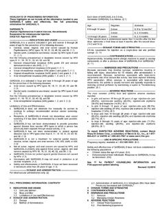
1 OPEN ACCESS ATLAS OF OTOLARYNGOLOGY, HEAD & neck OPERATIVE SURGERY MODIFIED & RADICAL neck DISSECTION Johan Fagan neck dissection removes potential or proven metastases to cervical lymph nodes. It is a complex operation and requires a sound knowledge of the 3-dimensional anatomy of the neck . Indications neck dissection may be elective (END) when done for clinically occult metastases, therapeutic (clinical metastases) or may be a salvage procedure (previously treated neck with surgery +/ radiation). END is indicated when the risk of having occult cervical nodal metastases exceeds 15-20%. Nodal Levels The neck is conventionally divided into 6 levels; Level VII is in the superior mediastinum (Figure 1). Figure 1: Classification of cervical nodal levels (Consensus statement on the classification and terminology of neck dissection. Arch Otolaryngol Head neck Surg 2008; 134: 536 8) Level I is bound by the body of the mandible above, the stylohyoid muscle posteriorly, and the anterior belly of the contralateral digastric muscle anteriorly.
2 The revised classification (Figure 1) uses the posterior margin of the submandibular gland as the boundary between Levels I and II as it is clearly identified on ultrasound, CT, or MRI. Level I is subdivided into Level Ia, (submental triangle) which is bound by the anterior bellies of the digastric muscles and the hyoid bone, and Level Ib (submandi-bular triangle). Level II extends between the skull base and hyoid bone. The posterior border of the sternocleidomastoid defines its posterior border. The stylohyoid muscle (alternately the posterior edge of the submandibular gland) defines its anterior border. The accessory nerve (XIn) traverses Level II obliquely and subdivides it into Level IIa (anterior to XIn) and Level IIb (behind XIn). Level III is located between the hyoid bone and the inferior border of the cricoid carti-lage. The sternohyoid muscle marks its an-terior limit and the posterior border of the sternocleidomastoid its posterior border.
3 Level IV is located between the inferior border of the cricoid cartilage and the clavi-cle. The anterior boundary is the sterno-hyoid muscle, and the posterior border is the posterior border of sternocleidomastoid. Level V is bound anteriorly by the posterior border of the sternocleidomastoid, and pos-teriorly by the trapezius muscle. It extends from the mastoid tip to the clavicle and is subdivided by a horizontal line drawn from the inferior border of the cricoid cartilage into Level Va superiorly, and Level Vb inferiorly. Level VI is the anterior, or central, compart-ment of the neck . It is bound laterally by the carotid arteries, superiorly by the hyoid bone, and inferiorly by the suprasternal notch. 2 neck Dissection Classification neck dissection operations are classified according to the cervical lymphatic regions that are resected (Figures 1, 2). Selective neck dissection (SND) is done for N0 necks (no clinical evidence of neck nodes) or for very limited cervical metasta-ses (Figure 2).
4 Central neck dissection encompasses only Level VI (Figure 1). Comprehensive or therapeutic neck dissec-tion involves surgical clearance of Levels 1-V and may either be a radical (RND) or modified (MND) neck dissection. RND includes resection of sternocleido-mastoid muscle (SCM) and accessory nerve (XIn) and internal jugular vein (IJV). MND preserves SCM and/or XIn and/or IJV. MND type I entails preservation of 1/3, usually XIn; MND type II entails preservation of 2/3, usually XIn and IJV; with MND type III all 3 structures are preserved. MND type II is most commonly done and is oncologically acceptable in the absence of adherence of cervical nodal metastases to XIn or IJV. Extended neck dissection includes addition-al lymphatic groups (parotid, occipital, Level VI, mediastinal, retropharyngeal) or non-lymphatic structures (skin, muscle, nerve, blood vessels etc.) not usually inclu-ded in a comprehensive neck dissection.
5 It has been proposed that neck dissections be more logically and precisely described and classified by naming the structures and the nodal levels that have been resected. (Ferlito A, Robbins KT, Shah JP, et al. Proposal for a rational classification of neck dissections. Head neck . 2011;33(3):445-50) Figure 2: Common types of neck dissection Modified neck dissection: Operative steps The detailed step-by-step description of neck dissection that follows refers to a right sided MND type I or II. RND involved the same surgical steps, other than that the IJV is double ligated superiorly and inferiorly with silk and with a silk transfixion suture passed through the vein, taking care not to include the vagus nerve (Xn) in the ligature. Anaesthesia, positioning and draping The operation is done under general anaes-thesia without muscle relaxation as eliciting movement on mechanical or electrical stim-ulation of the marginal mandibular, hypo-glossal (XIIn) and accessory nerves assist with locating and preserving these nerves.
6 It is a clean operation unless the upper aero-digestive tract is entered, and antibiotics are therefore not required. With an experienced surgeon, blood transfusion is rarely re-quired. The patient is placed in a supine position with the neck extended and turned to the opposite side. Surgical draping must allow monitoring for movement of the lower lip 3 with irritation of the marginal mandibular nerve, and must provide ACCESS to the clavicle inferiorly, the trapezius muscle posteriorly, the tip of the earlobe superiorly and the midline of the neck anteriorly. The drapes are sutured to the skin. Incisions and flaps Incisions should take into consideration ACCESS that may be required to resect the primary tumour, cosmetic factors, and the blood supply to the flaps. Flaps are elevated in a subplatysmal plane with a knife or with monopolar electrocautery. Making the flaps too thin may compromise the blood supply to the skin flaps.
7 Figure 3 demonstrates incisions commonly used for MND done in association with cancers of the oral cavity, oropharynx, nasal cavity sinuses and skin cancers of the midface. The transverse skin incision can be extended across to the opposite side with bilateral neck dissections or can be exten-ded superiorly to split the lower lip in the midline to gain ACCESS to the oral cavity. Care should be taken not to place the trifurcation of the incision over the carotid artery, as skin loss at this point may expose the carotid artery with its attendant risks. Figure 4 demonstrates the hockey stick incision. This can be extended into a pre-auricular skin crease and is particularly useful for combined parotidectomy and neck dissection. It has the advantage that the scar of the vertical limb along the anterior border of the trapezius can be hidden by long hair. Care must be taken in patients who have been previously irradiated as the posteroinferior corner has a tenuous blood supply and may slough, having to heal by secondary intention.
8 Figure 3: Incisions for neck dissection combined with oral cavity cancer resection Figure 4: Hockey-stick incision for neck dissection combined with parotidectomy With laryngectomy patients, MND can be done either via a wide apron flap, or by lateral extensions from the apron flap (Figures 5, 6). 4 Figure 5: Wide apron flap Figure 6: Apron flap with lateral extensions neck dissection: Operative steps Figure 7 shows a completed right-sided MND type II. The superimposed numbers indicate the sequence of the main operative steps that will be referred to in the description of the surgery that follows. Step 1 (Figure 7) The neck is opened via a horizontal incision placed in a skin crease at about the level of the hyoid bone. The incision is made through skin, subcutaneous fat, and platys-ma muscle. Identify the external jugular vein and greater auricular nerve overlying the sternocleidomastoid muscle (SCM) (Figure 8).
9 Figure 7: Completed right MND type II with sequence of operative steps Figure 8: Note platysma muscle (transec-ted), and the external jugular vein and greater auricular nerve overlying the SCM Next the superior flap is elevated with cautery until the submandibular salivary gland is identified. The submandibular gland fascia is then incised inferiorly over the gland to avoid injury to the marginal mandibular nerve (Figure 9). 5 Figure 9: Incision of submandibular saliva-ry gland capsule The surgeon then resects the fat and lymph nodes from the submental triangle (Level Ia). A subplatysmal dissection of the over-lying skin is extended to the opposite anterior belly of digastric muscle, taking care not to injure the anterior jugular veins. The submental triangle is resected inferiorly to the hyoid bone with electrocautery. The deep plane of dissection is the mylohyoid muscles (Figures 10 & 11).
10 Figure 10: Resection of submental triangle Figure 11: Resection of submental triangle onto mylohyoid muscles Step 2 (Figure 7) The surgeon next addresses Level Ib of the neck . Because the marginal mandibular nerve runs in an extracapsular plane, the submandibular gland capsule is dissected from the gland in a superior direction in a subcapsular plane (Figure 9). The marginal mandibular nerve does not need to be routinely identified. The assistant however watches for twitching of the lower lip, as this indicates proximity of the nerve. The facial artery and vein are identified by blunt dissection with a fine haemostat (Figure 12). The marginal mandibular nerve crosses the facial artery and vein (Figure 12). Next attention is directed at the fat and lymph nodes tucked anteriorly and deeply between the anterior belly of digastric and mylohyoid muscle. These nodes are espe-cially important to resect with malignan-cies of the anterior floor of mouth.