Transcription of Opercular syndrome: Case reports and review of …
1 145 Opercular syndrome : case reports and review of literatureThapa Lekhjung, Paudel Raju, Rana PVSD epartment of Neurology, College of Medical sciences-Teaching Hospital, Bharatpur, Chitwan, NepalAbstract Five cases of bilateral Opercular syndrome of vascular etiology are reported. Cortical pseudobulbar palsy ( spastic anarthria and inability to swallow) with dissociation of automatic voluntary movements in the affected muscles are the essential features of this syndrome . Additional motor and sensory symptoms differentiate its subtypes. All 5 patients had bilateral Opercular syndrome . The unusual features was its occurrence as the presenting feature of preeclampsia in a young lady, and the development of the transient syndrome following a right focal seizure with generalization at high altitude in a young female trekker who had an old unilateral infarct in left Opercular region.
2 Whereas the limb motor weakness recovered well, the recovery was unsatisfactory for speech and for Asia 2010; 15(2) : 145 152 Address correspondence to: Dr Thapa Lekhjung, Department of Neurology, College of Medical sciences-Teaching Hospital, Bharatpur, Chitwan, NepalINTRODUCTIONThe Opercular syndrome is a rare disorder due to bilateral lesions of Opercular cortex surrounding the insula, which is separated by the ascending and the posterior rami of the lateral sulcus into (a) Frontal operculum formed by posterior part of the inferior frontal gyrus ( pars-triangularis, pars-opercularis and even by the caudal portion of the pars-orbitalis); (b) Fronto-parietal opercula formed by the lowermost part of the precentral and postcentral gyrus and the anterior and lowermost part the inferior parietal lobule.
3 And (c) Temporal opercula formed by the superior temporal The syndrome was fi rst described by Magnus in 18372 and is also known as Foix-Chavany-Marie syndrome (FCMS)3, facio-labio-glosso-pharyngo-laryngo-brac hial paralysis or cortical type of pseudobulbar Further, it is classifi ed based on the site of lesion as bilateral anterior Opercular syndrome (lesion in both anterior or frontal operculum)1,5, Opercular -subopercular syndrome (lesion in Opercular cortex one side and the subopercular lesion on the other side)6, subopercular syndrome (lesions in subcortical corticobulbar projections only)4,7, unilateral anterior syndrome involving frontal operculum1,4, and posterior syndrome involving frontoparietal ,8 Its manifestations includes central type of facial paresis, diffi culty in opening of mouth, chewing the food, protrusion of tongue and inability to speak and swallow.
4 Additional features include trismus9, ageusia10 and pseudo-ophthalmoplegia ( ptosis with weakness of conjugate gaze to opposite side and deviation of head and eyes to the side of lesion). In addition unilateral anterior syndrome is reported involving frontal operculum and presenting with contralateral and upper limb paresis and inability to speak (Piere Marie s anarthria).1 It differs from bulbar palsy by preservation of jaw jerk, pharyngeal refl ex and by the absence of fasciculation, atrophy and phenomenon of denervation and unlike pseudobulbar palsy the pathological laughter and emotional disturbances are lacking. The etiology in most of the reported cases is vascular (thrombosis or embolism) involving branches of middle cerebral artery supplying the Opercular ,5 Other lesions producing the syndrome include astrocytoma11, developmental bilateral perisylvian cortical dysplasia12,13, herpes simplex encephalitis14-16, progressive supranuclear motor system degeneration17, bilateral toxoplasmosis in AIDS18, and in multiple Weller et al20 reviewed 62 cases and classifi ed Opercular syndrome based on etiology; (a) Classical form most often related to vascular etiology; (b) Subacute form due to central nervous system infections.
5 (c) Developmental form most often related to neuronal migration disorders; (d) Reversible form in children with epilepsy; and (e) Rare type related with neurodegenerative disorders. Some authors consider Foix-Chavany-Marie syndrome in children a different We report this rare syndrome with some previously unreported observations with review of the AsiaAugust 2010146 case reports Patient 1A 35 years old lady, a resident of , while trekking in Nepal at height of 3,000-4,000 meters, had a right focal seizure with secondary generalization lasting for 10-15 minutes. Following this episode she developed diffi culty in swallowing and inability to speak and mild weakness on the right half of the body predominantly affecting the upper limb.
6 She had an ischemic stroke 2 years back, manifesting with right hemiplegia and non-fl uent aphasia. She was diagnosed as left middle cerebral artery infarction with no evident risk factors for stroke. She was treated conservatively with aspirin and made full neurological recovery. At admission she was afebrile, with BP 140/90 mms Hg, pulse 64/minute. Neurological examination revealed normal higher mental functions. She could not vocalize but, she could follow spoken and written command and could copy normally. She could recognize and name the objects. She could not open the mouth or protrude the tongue or swallow. However, opening of mouth was noted during yawning and laughing. She also had facial paresis of central type, sluggish palatal and pharyngeal refl exes, pronator drift in right arm, brisk deep tendon refl exes and extensor plantar response on the right side.
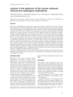
7 Fundus and other examinations were normal. Hematological and biochemical parameters, ECG and echocardiography were normal. EEG revealed focal slowing and epileptic discharges from left frontotemporal region. CT scan (Figure 1) revealed old Opercular infarcts in left Opercular and sub Opercular areas with ipsilateral dilatation of lateral ventricles. Carbamazepine was added to aspirin. When transferred back to after 7 days, she had marked improvement in her speech but only partial improvement of 2A 24 years old lady, a primigravida with 6 months history of amenorrhea, was admitted with acute neurological illness manifesting with moderately severe global headache and inability to speak.
8 Figure 1: Brain CT scan of Patient 1 showing infarcts in left Opercular and subopercular region147 There was no history of loss of consciousness, vomiting, diplopia, seizures, transient ischaemic attack or of a similar episode in the past. She denied history of trauma, fever or vaccination, diabetes mellitus and hypertension. She was not a booked case and had not attended antenatal clinic services. At admission she was afebrile with pulse of 110/min, BP 180/124 mm Hg, respiration 24/min and had mild bilateral pitting edema. No icterus, cyanosis or lymphadenopathy were detected. She was conscious and responding to commands. However, she could not vocalize. She could not open her mouth, protrude her tongue or swallow.
9 Involuntary movements were preserved. She had central facial paresis, mild hemiparesis (upper limb affected more than lower limb) and brisk refl exes on right side and extensor plantar response on both sides. All other systems including the cardiorespiratory system were normal. The fundal height was commensurate with the period of amenorrhea. Evaluation for risk factors for ischemic stroke was negative. Except for slight albuminuria and mild rise in hepatic enzyme, other liver functions, hemogram, metabolic parameters, coagulation profi le, plasma fi brinogen level, ECG and ultrasonography of abdomen were normal. Antinuclear factor and tests for antiphospholipid antibodies were negative.
10 CT scan (Figure 2) revealed large bilateral cortical infarct in the posterior frontal region involving both Opercular areas. She was treated as a case of severe preeclampsia with magnesium sulphate, antihypertensive drugs (calcium channel blockers), aspirin, anticerebral edema measures and termination of pregnancy. Anticonvulsant was added later for seizure prophylaxis. She improved gradually and when discharged after three weeks, she could speak a few syllables but the swallowing did not improve. Patient 3A 75 years old lady, a known case of atrial fi brillation, presented with one day history of weakness right half of the body (upper limb more than lower limb), inability to speak and swallow.