Transcription of Patellar Femoral Pain Syndrome (PFPS) - ou h
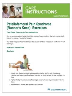
1 Patellar Femoral Pain Syndrome (PFPS). Advice and management OxSport Department of Sport and Exercise Medicine This booklet has been written to help guide you through the management of your Patellar Femoral Pain Syndrome . It is important that you read this booklet, so you have a better understanding of the condition and its management. What is Patellar Femoral Pain Syndrome (PFPS). PFPS is a common condition causing knee pain in both athletes and non-athletes, which can affect both men and women of all ages. PFPS occurs due to overloading of the front of the knee, behind the kneecap. It is more common in people who take part in sport, particularly those whose sports involve running or jumping, such as tennis, football or badminton. Sometimes you will hear this condition called anterior knee pain, chondromalacia patellae or patellofemoral disorder, but these mean the same thing as PFPS. page 2. What causes PFPS? PFPS most often occurs without an injury to the knee, but can sometimes be a result of an injury, such as a fall onto the knee.
2 If you have not injured your knee, it may be difficult to find one specific cause of your PFPS, as it usually occurs due to a combination of factors. It is often associated with the start of a new activity, an increase in the intensity and/or frequency of an existing activity, or following a period of reduced activity that leads to weakening of the muscles. There are many contributing factors, which can vary from person to person, but it is rare that are they all present. The diagram below shows some of the main factors which can contribute to the development of PFPS. Factors contributing to the development of PFPS. Tight muscles ( quadriceps). Poor foot stability Abnormal Poor hip control biomechanics Tight iliotibial band Quadriceps dysfunction Training errors ( Abnormal kneecap sudden change in movement running distance). Excessive patellofemoral joint pressure Equipment errors ( wrong footwear) PAIN. page 3. What are the symptoms of Patellar Femoral Pain Syndrome ?
3 Pain is the main symptom of PFPS. It can be felt anywhere around the kneecap and even at the back of the knee. It can affect one or both knees. There are a variety of other symptoms that people with PFPS. experience, including: clicking or clunking mild swelling a feeling of instability (like your knee might give way). pain when squatting or coming downstairs discomfort or pain when sitting for long periods (such as going to the cinema or taking long airplane journeys). X-rays and scans We do not routinely use X-rays or scans (imaging) for the diagnosis of PFPS, as looking at your medical history and examining your knee is usually enough for us to make the diagnosis. If we do need to carry out imaging, this may be an ultrasound scan, X-ray, or MRI scan, to exclude other causes of knee pain. MRI scans may show changes behind your kneecap (such as fraying of the cartilage) that are common in people both with and without PFPS. Findings like these often do not relate to your PFPS.
4 Please discuss any findings from the scan with the doctor who requested the scan for you. They will let you know if the results of the scan are important or not. page 4. Treatment A structured exercise programme is the best treatment for your PFPS. Research shows this to be the best therapy. Before you begin your exercise programme, your doctor or physiotherapist will examine you. They will identify any particular problems you might have with specific muscle groups. They will also look at how well you are moving and recommend some specific exercises for you to do as a home exercise programme'. You may also be referred for a course of physiotherapy, if you need someone to work more closely with you during your rehabilitation . If this is the case, your physiotherapist may use additional techniques to help with your rehabilitation , such as using tape which is stuck to your skin to help support your knee, or soft tissue techniques. Pain can sometimes stop you from rehabilitating properly with PFPS, because it can make it very hard to train your muscles effectively.
5 As well as regular painkillers such as paracetamol and ibuprofen, we may suggest you take other sorts of painkillers to help control your symptoms better and enable you to strengthen your muscles. You may also be referred to an orthotist, who specialises in assessing your foot biomechanics (how the bones, muscles and tendons move). They may prescribe shoe inserts called orthotics'. These will help change your foot posture, if this is contributing to your PFPS. page 5. HOME EXERCISE PROGRAMME. This home exercise programme is divided up into specific sections, each addressing different issues that can cause PFPS. It includes: stretches strengthening balance exercises core stability (strengthening the muscles around your tummy and pelvis). plyometrics (powerful movements, such as jumping). Sometimes, while doing the exercises, you may feel an increase in your symptoms around your knee cap. This is normal and you will find that as you progress with your exercises your symptoms will improve.
6 You may not feel the benefits of the exercises until approximately 3 months after starting, so it is important that you continue with the programme. page 6. Stretches Hold for 1 minute on each muscle group. When stretching you should not push the stretch into pain. You should instead feel a strong stretching sensation, which should ease off when you stop. You should hold a continuous stretch and not bounce at the end of the movement. The following programme is designed to help with flexibility of the whole leg. Gastrocnemius muscle stretch (right leg stretch shown in picture). Step forwards with your left leg, leaving your right leg behind you and straight. Lean forwards, while keeping your right heel on the floor. You should feel a stretch at the back of your right calf muscle. Hold for one minute, then slowly release and switch legs. page 7. Soleus muscle stretch (right leg stretch shown in picture). Prepare as for the previous stretch (gastrocnemius), but do the stretch with your right knee bent, not straight.
7 You should feel a stretch in the lower part of your right calf muscle. Hold for one minute, then slowly release and switch legs. Quadriceps muscle stretch (right leg stretch shown in picture). Bend your right knee and grab hold of your right ankle with your left hand. Make sure your knees are together, then push your right hip backwards. You should feel a stretch at the front of your right leg. Hold for one minute, then slowly release and switch legs. page 8. Gluteus stretch (right leg stretch shown in picture). Lying on your back, place your right foot against a wall in a relaxed position Raise your right leg and place your right ankle onto your left knee. You should feel a stretch in your right buttock. To increase the stretch, move closer to the wall. Hold for one minute, then slowly release and switch legs. Hamstring stretch (right leg stretch shown in picture). Lying on your back, raise your right leg and hold behind your knee with both hands. In this position, straighten your right knee as far as it will go.
8 You should feel a stretch at the back your right leg. Hold for one minute, then slowly release and switch legs. page 9. Iliotibial band soft tissue release, using a tennis ball or foam roller Lie on your side with your tennis ball or foam roller under the middle of the outside of your thigh. Move your body up and down, rolling the ball or roller along the outside edge of your leg. Do this for 30. seconds to 1 minute on each leg. page 10. Strengthening exercises Wall slides Stand with your back against the wall and feet shoulder width apart, about a foot away from the wall. Slide down the wall, keeping your shoulders back until you start to feel any discomfort, then slide back up the wall. Gradually build up to a knee bend of 90 (a right angle). When you can achieve a 90 . knee bend, you can add in squeezing a ball in between your knees whilst doing this exercise. Forward lunge For this exercise your front leg should be your affected leg. Starting in a standing position, step forward into a lunge position, as shown in the picture.
9 Keep your body upright and do not let your leading knee go beyond your toes. The knee of your trailing leg should not be touching the floor. page 11. Proprioception (balance awareness). Stand on your affected leg for 1 minute, without putting your foot down, to challenge your balance. If you are able to do this for 1 minute, try to do the same exercise with your eyes shut for 1 minute. If you are able to do this, then progress to standing on an unstable surface (such as a folded towel). Make sure at all times that you are in a safe environment, so you do not lose your balance and injure yourself. page 12. Core stability Core stability concentrates on strengthening the muscles around your pelvis and tummy. You need good muscular control of your pelvis, as this acts as the stable base from which your legs move. Without a stable pelvis, the forces going through your legs can be increased and unpredictable, which can lead to injury. To engage your deep tummy muscles (transverse abdominus), think about pulling your belly button inwards.
10 Imagine that you are trying to do up a pair of trousers that are a size too small. While doing this, do not hold your breath! You should feel your tummy muscles tighten slightly. You can feel this if you place your fingers over the lower part of your tummy. When doing the core stability exercises, make sure you engage your tummy muscles before doing each exercise. Lower and lift Lying on your side, engage your tummy muscles. Raise your uppermost leg approximately 6 inches off the ground, then lower it again to the start position. While moving your leg, do not let your pelvis move. Repeat 50 times, then switch to the other side. page 13. Bridging level 1. Lie on your back with your knees bent. Engage your tummy muscles. Tilt your pelvis backwards, as if pressing your lower back into the floor. Squeeze your bottom muscles together. Slowly lift your pelvis and back off the floor to the bridge position shown. Hold for 5 seconds and then slowly return back to the starting position.
