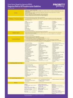
Transcription of Peptic Ulcer Disease: Introduction - Hopkins Medicine
1 Figure 1. Location of the stomachand duodenum in the body. [ ] Peptic Ulcer disease : Introduction Peptic Ulcer disease represents a serious medical problem. Approximately 500,000 new cases are reported each year, with 5 millionpeople affected in the United States alone. Interestingly, those at the highest risk of contracting Peptic Ulcer disease are thosegenerations born around the middle of the 20th century. Ulcer disease has become a disease predominantly affecting the olderpopulation, with the peak incidence occurring between 55 and 65 years of age. In men, duodenal ulcers were more common thangastric ulcers; in women, the converse was found to be true. Thirty-five percent of patients diagnosed with gastric ulcers will sufferserious complications. Although mortality rates from Peptic Ulcer disease are low, the high prevalence and the resulting pain,suffering, and expense are very can develop in the esophagus, stomach or duodenum, at the margin of a gastroenterostomy, in the jejunum, in Zollinger-Ellison syndrome, and in association with a Meckel's diverticulum containing ectopic gastric mucosa.
2 Peptic Ulcer disease is one ofseveral disorders of the upper gastrointestinal tract that is caused, at least partially, by gastric acid. Patients with Peptic Ulcer diseasemay present with a range of symptoms, from mild abdominal discomfort to catastrophic perforation and bleeding. What is Peptic Ulcer disease ?Gastric and duodenal ulcers are breaks in the gastric and duodenal mucosa. Both gastric and duodenal ulcers relate to the corrosive action of pepsin andhydrochloric acid on the mucosa of the upper gastrointestinal tract. Ulcers generally range between 3 mm and several centimeters in patients with Peptic Ulcer disease present with abdominal discomfort, pain or nausea. The pain is located in the epigastrium and usually does not , these symptoms are neither sensitive nor specific.
3 Pain radiating to the back may suggest that an Ulcer has penetrated posteriorly, or the pain may bepancreatic in origin. Pain radiating to the right upper quadrant may suggest disease of the gallbladder or bile may describe the pain of Peptic Ulcer as burning or gnawing, or as hunger pains slowly building up for 1 2 hours, then gradually decreasing. Use of antacidsmay provide temporary relief. Classically, gastric Ulcer pain is aggravated by meals, whereas the pain of duodenal ulcers is relieved by meals. Hence, patients withgastric ulcers tend to avoid food and present with weight loss, while those with duodenal ulcers do not lose weight. It is important to remember that although thesepatterns are typical, they are not pathognomonic. The nature of the presenting symptoms alone does not permit a clear differentiation between benign ulcers andgastric neoplasm.
4 Copyright 2001-2013 | All Rights North Wolfe Street, Baltimore, Maryland 21287 Peptic Ulcer disease : Anatomy AnatomyThe stomach is located in the upper part of the abdomen just beneath the diaphragm (Figure 1). The stomach is distensible and on a free mesentery, therefore, thesize, shape, and position may vary with posture and content. An empty stomach is roughly the size of an open hand and when distended with food, can fill much ofthe upper abdomen and may descend into the lower abdomen or pelvis on standing. The duodenum extends from the pylorus to the ligament of Treitz in a sharpcurve that almost completes a circle. It is so named because it is about equal in length to the breadth of 12 fingers, or about 25 cm. It is largely retroperitoneal and itsposition is relatively fixed.
5 The stomach and duodenum are closely related in function, and in the pathogenesis and manifestation of stomach may be divided into seven major sections. The cardia is a 1 2 cm segment distal to the esophagogastric junction. The fundus refers to the superiorportion of the stomach that lies above an imaginary horizontal plane that passes through the esophagogastric junction. The antrum is the smaller distal one-fourth toone-third of the stomach. The narrow 1 2 cm channel that connects the stomach and duodenum is the pylorus. The lesser curve refers to the medial shorter border ofthe stomach, whereas the opposite surface is the greater curve. The angularis is along the lesser curve of the stomach where the body and antrum meet, and isaccentuated during peristalsis (Figure 2).
6 Figure 2. A, Normal anatomy of the stomach and duodenum; B-D, corresponding endoscopic images. The duodenum extends from the pylorus to the ligament of Treitz in a circle-like curve and is divided into four portions. The superior portion is approximately 5 cm inlength, beginning at the pylorus, and passes beneath the liver to the neck of the gallbladder. The first part of the superior portion (2 3 cm) is the duodenal bulb. Thedescending or second part of the duodenum takes a sharp curve and goes down along the right margin of the head of the pancreas. The common bile duct and thepancreatic duct enter the medial aspect of this portion of the duodenum at the major papilla either separately or together. The duodenum turns medially, becoming thehorizontal portion, and passes across the spinal column, inclining upward for 5 8 cm.
7 The ascending portion begins at the left of the spinal column, ascending left ofthe aorta for 2 3 cm, and ends at the ligament of Treitz, where the intestine angles forward and downward to become the jejunum. Copyright 2001-2013 | All Rights North Wolfe Street, Baltimore, Maryland 21287 Peptic Ulcer disease : Causes Protective vs. Hostile Factors No gastric acid, no Peptic Ulcer is a misconception. Excessive gastric acid secretion is only one factor in the pathogenesis of Peptic Ulcer disease . Decreasedmucosal defense against gastric acid is another cause. The integrity of the upper gastrointestinal tract is dependent upon the balance between hostile factors suchas gastric acid, H. pylori, NSAIDs and pepsin, and protective factors such as prostaglandins, mucus, bicarbonate, and blood flow to mucosa affectinggastrointestinal mucosa (Figure 3).
8 Figure 3. A, Protective factors; B, hostile to gastric and duodenal mucosa develops when deleterious effects of gastric acid overwhelm the defensive properties of the mucosa. Inhibition of endogenousprostaglandin synthesis leads to a decrease in epithelial mucus, bicarbonate secretion, mucosal blood flow, epithelial proliferation, and mucosal resistance to mucosal resistance increases the incidence of injury by endogenous factors such as acid, pepsin, and bile salts as well as exogenous factors such as NSAIDs,ethanol and other noxious agents (Figure 4).Figure 4. Pathogenesis of Peptic Ulcer disease . Helicobacter pyloriH. pylori is the etiologic factor in most patients with Peptic Ulcer disease and may predispose individuals to the development of gastric carcinoma.
9 H. pylori colonizesin the human stomach (Figure 5). The method of H. pylori transmission is unclear, but seems to be person-to-person spread via a fecal-oral route. The prevalence ofH. pylori in adults appears to be inversely related to the socioeconomic status. It is also thought that water is a reservoir for transmission of H. 5. A, H. pylori resident on the gastric epithelium; B, electron micrograph. Nonsteroidal Anti-Inflammatory Drugs (NSAIDS)A small but important percentage of patients have adverse gastrointestinal events associated with NSAID use that results in substantial morbidity and mortality. Riskfactors for the development of NSAID-associated gastric and duodenal ulcers include advanced age, history of previous Ulcer disease , concomitant use ofcorticosteroids and anticoagulants, higher doses of NSAIDs, and serious systemic disorders.
10 The concept of gastroduodenal mucosal injury has evolved from thenotion of topical injury to concepts that involve multiple mechanisms. NSAIDs initiate mucosal injury topically by their acidic properties. By diminishing the hydrophobicity of gastric mucus, endogenous gastric acid and pepsin may injuresurface epithelium. Systemic effects of NSAIDs appear to play a predominant role through the decreased synthesis of mucosal prostaglandins. The precursor ofprostaglandins, arachidonic acid, is catalyzed by the two cyclo-oxygenase isoenzymes, cyclo-oxygenase-1 and cyclo-oxygenase-2. The gene for cyclo-oxygenase-1,the housekeeping enzyme, maintains the homeostasis of organs. Cyclo-oxygenase-2, the inflammatory enzyme, is inducible. Although NSAIDs can inhibit bothpathways, only the gene for cyclo-oxygenase-2 contains a corticosteroid-responsive repressor element (Figure 6).