Transcription of Physiology Respiration Doppler Veins - msecho.org
1 Respiration : Effects Physiologic effects of Respiration are secondary to How to Utilize Respiratory Manouvers in intra thoracic and intra abdominal pressure in systemic and pulmonary venous return in Echocardiography pericardial pressure constraint Practical Applications in Daily Practice interdependence Karthik Ananthasubramaniam, MD FACC FASE FASNC. Associate Professor of Medicine Anatomic effects of Respiration are due to Wayne State University Director, Echocardiography and Nuclear Cardiology/PET Laboratory of lungs and interference Henry Ford Hospital, Detroit MI. out of lateral shadows translation of the diaphragm Disclosures Research grant support American Society of Echocardiography Astellas Pharma Global Development, Inc CV Therapeutics GE Healthcare Molecular Insight Pharmaceuticals Speakers Bureau/Honoraria : Astellas Pharma Global Development, Inc Consultant : Lantheus Medical Imaging Therefore diastolic filling pressure No conflicts of interest for this talk is the result of LV stiffness and intra pericardial pressure Respiration and Image Quality Objectives With inspiration : in AP diameter inflation Less heart available for imaging and posterior motion of heart Outline effects of Respiration on heart imaging is an exception.
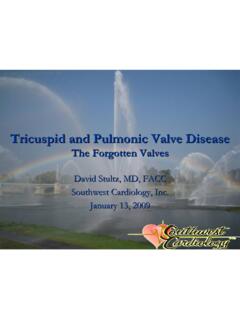
2 Heart images clearer with inspiratory diaphragm descent Outline how Respiration can affect 2d, Doppler imaging Tips for better accquisition Understand role of Valsalva manouver in echo With expiration : Use of respiratory in different cardiac conditions 1. Lung deflation ( particularly in steep left lateral position with left arm up). Physiology of Respiration Respiration and Doppler of Veins Ventricular Interdependence Hepatic vein Doppler pulmonary vein Doppler Sig inspiratory increase of S and D Minimal resp changes in S. SVC flow mimics HV Doppler changes but d and VTId increase exp Respiration and echo Resp and pulmonary Venous flow Measurements s and VTIs not affected d and VTId increase with exp Inspiratory decrease in LV end diastolic dimensions as measured by M mode Hence s/d < 1 and VTIs/VTId < 1 in exp This may sometimes explain discrepancies between centerline PSAX measurements and those obtained by M mode Mechanisms in health Possible reasons: Left right interactions moves medially during inspiration= ?
3 Tangential M mode cut Diastolic dysfunction causes higher LV diastolic in LV volumes in inspiration pressures ( preload reduction and increase afterload or impedance to LV emptying ) So intra cardiac interactions of septa in resp decreased How to measure : suspended quiet Respiration Insp Exp make sure to instruct patients to avoid So d and VTId do not vary Valsalva during held expiration as it degrades with insp and exp image quality Lack of variation a clue Respiration and Doppler of AV and Systemic Valves Respiration And Doppler Issues MV TV Fixed Doppler sampling errors can occur during Respiration due to change in heart position and diaphragmatic traction Mitr Tricus Parallel positioning and avoidance of angle of incidence issues is key. Less than 10% error is most preferable given the quadratic relationship of pressure and velocity Transmitral v.
4 Varies by <10%, Tricuspid v. varies by <20%, increasing with exp. increasing with insp. End expiration apnea most preferred for Doppler sampling AV PV. Similar sampling issues affect tissue Doppler Aortic Pulm Respiration , IVC and echo RAP: Respiration and Tissue Doppler ? Time for Reappraisal Brennan et al : JASE 2007. RAP measured from IVC in subcostal view and compared to RHC. This study questions using tight cutoffs of 5 mm to define RAP based on IVC. Overall IVC size 2cm served as the most optimal cutoff for RAP > or < 10 ( sens 73% spec 85% ). IVC collapsibility index of 40% was the best predictor for response ( sens 74% spec 84%). During normal Respiration During end expiratory apnea Sample volume can move with Respiration . Best obtained with held expiration Respiration and IVC A New Classification for RAP.
5 Minimal size observed in end inspiration Influenced by patient position : Largest in right lateral position Intermediate in supine position Smallest in left lateral position Normal ranges : 1 2 cm ( upper normal range ASE guidelines cm ). Can be large and minimally reactive in young patients, athletes can have large IVC and normal collapsibility index Sniff test captured in real time and m mode 3 5 loop rhythm capture will be helpful . M mode cursor to be placed away from IVC RA junction Brennan et al : JASE 2007. Respiration and IVC Respiration and SVC. IVC Diameter % Collapse with Sniff Estimated CVP. < cm Full collapse 0 mmHg cm >50% collapse 1-5 mmHg > cm >50% collapse 6-10 mmHg > <50% collapse 11-15 mmHg > cm 0% collapse >16 mmHg Dilated IVC in vented patients does not mean elevated RAP.
6 Collapsed IVC ( >50% ) indicates dehydration in vented patients Normal : Systolic forward flow = RA relaxation during RV contraction Specificity for predicting RAP increases when IVC size is measured at end expiration by M Diastolic forward wave = rapid RV filling wave mode AND at end disatole Vary with Respiration with both waves higher during inspiration than expiration Mechanism of Flow Changes in SVC. Possible clue to Pul HTN in COPD Valsalva in Diastolic Function a. Need to instruct patients correct how to do it. To avoid forceful SVC is an intra thoracic structure : Interrogation is from right supraclavicular fossa with deep inspiration prior to the forced expiration. Doppler PW sample volume of 2mm. b. Forced expiration against closed nose and mouth. Changes in RA pressure following change in pleural pressure causal SVC flow variations c.
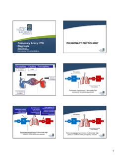
7 Useful to clarify pseudonormal mitral inflow patterns. Expiratory systolic flow in SVC is a measure of RA flow reserve d. In normal patients if the E wave drops by 20cm/sec that is a good effort useful to In Pul HTN : RV filling is restricted RA pressure is elevated and SVC forward variation is interpretation. E/A ratio remains above 1 despite the change in patients with normal blunted diastolic function In Pul HTN : RA preload reserve is enhanced to compensate for RV dysfunction and hence SV expiratory forward flow velocity is increased e. A E wave drop > 50 cm/sec is highly specific ( 100% spec) for elevated filling pressures accompanied by reversal to impaired relaxation pattern to confirm Hence SVC interrogation would be a useful adjunct in patients with significant lung disease pseudonormal pattern although lower levels may also where TR jet is not well seen or minimal to no TR is present precluding good assessment of PASP indicate the same diagnosis SVC Doppler Index and Pul HTN The Valsalva Manouever : Its Effects on Intracardiac Volumes Baseline Valsalva The Valsalva Manouver Phase 1 start strain : increased BP , transient increase venous return Phase 2 strain maintained : decrease BP, pulse pressure and sinus tachycardia Phase 3 strain release : progressive fall in BP.
8 Phase 4 post release phase : overshoot of BP and reflex bradycardia Correlations between LVEDP and the baseline E/A ratio (A), the change of the E/A ratio during Valsalva (B), And the change of the A wave during Valsalva (C). Patients with a baseline E/A ratio of <1 (impaired relaxation pattern) despite substantially elevated LVEDP are represented by the gray circles. Schawammenthal et al AJC 2000. Relaxation and Compliance Tug of War As diastolic dysfunction progresses : 2 scenarios can play out or co exist Impaired relaxation gives way to increased LV stiffness ( decreased compliance ). Severely impaired relaxation persist despite progressive worsening of compliance Normal diastology Normal filling pressures Impaired relaxation Is It This or That ? Normal filling pressures Beat to beat variation of E/A with Respiration E/A < 1 in Insp E/A > 1 Exp DT shorter in Exp than Insp Impaired relaxation Elevated filling No effects of resp on A pressures wave Pseudonormal diastology This pattern associated with mpaired tissue Doppler Could be a clue to diastolic dysfunction Respiration and PFO Detection Detection of PFO.
9 TEE considered highly sensitive even more than TCD SPARC Study Protocol No shunt small shunt 2 rest injections 2 injections with cough Moderate shunt large shunt 2 injections with Valsalva TEE PFO and Respiration Cough or Valsalva is widely employed Both false negatives and false positives can occur related to the respiratory effort False negative : poor effort in combination with sedation or lack of understanding of how the Valsalva is to be done Solution : instruct pt prior to TEE. A good idea would be to perform a bubble study with Valsalva prior to sedation and intubation both as a practice run and as check to pt compliance and understanding of what is expected False positive : Spurious contrast misinterpreted as shunt . small pulm AV malformation causing bubble appearance Detection of PFO Spurious contrast : Snow Storm Has been described with cough and Valsalva maneuver Methodologic Issues Has been described with TTE but most often TEE.
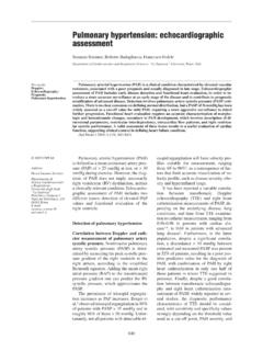
10 Number of injections Valsalva blood pooling in pulmonary Veins rouleax formation Valsalva Valsalva Use of maneuvers Coughing Coughing Release of Valsalva drops LA pressure rush of pulmonary vein blood into LA. Femoral approach Use of color Doppler Saline Bubbles Spurious contrast Spontaneous spurious bright and localized onset faint and weak intensity Contrast likley due the combination of rouleaux and Harmonic imaging (TTE) cavitation as blood enters LA. Stronger echodensity Weaker echodensity after Valsalva release Transmitral Doppler Persistent evanescent Spurious Contrast, SEC and Respiration Detection of PFO. SEC ( Spontaneous echo contrast ) versus Spurious contrast Causes of False Positive TEE. Low flow states No specific condition Spontaneous occurrence ** Only with cough /. valsalva Transpulomonary microbubbles Gain dependent Gain dependent Solution: for exit of bubbles with Valsalva release from septum or pulmonary vein.