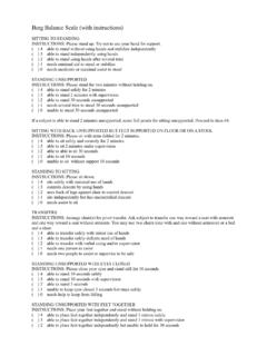
Transcription of Physiotherapy in Burns, Plastics and Reconstructive Surgery
1 Impairment and Disability Short Course 19 April 2013 Aoife Hale Rhona O Donovan Sarah Diskin Sarah McEvoy Claire Keohane Geraldine Gormley PY4017/4019 Module Leader: Norelee Kennedy Physiotherapy in Burns, Plastics and Reconstructive Surgery ii Preface This booklet is the culmination of our research in relation to best practice for Physiotherapists working in the area of Burns, Plastics and Reconstructive Surgery . It should be used in tandem with our presentation. It is not an exhaustive source, and should be used in conjunction with the referenced materials and any new research which may emerge in the future. It is not intended as a replacement for clinical reasoning, but to aid you in the process of your assessment and treatment planning and execution.
2 Acknowledgements We would like to thank the following specialists who contributed their guidance and knowledge in the completion of this project Catherine O Sullivan, Senior Physiotherapist UCHG, Alesha Kelly, Senior OT, UCHG, Fionnuala Cassidy, Senior Occupational Therapist in Burns, St James Hospital Dublin, and Kirstin Aschbacher, Senior Psychologist University College San Francisco. We are grateful for the guidance and advice provided by Norelee Kennedy, our module leader and to Oliver McGarr whose new perspective and guidance was invaluable in the creation of our presentation. Finally, thank you to Brian Stewart for his help in editing our video case study.
3 Iii Contents Acknowledgements .. ii Part 1 2 Section 1: Introduction .. 3 Epidemiology .. 3 Mechanism of Injury .. 3 Review of the Skin .. 4 Types of Burn .. Error! Bookmark not Physiology of Burns .. 6 Tissue Healing .. 9 Burn Associated Pain .. 13 Section 2: Burn Assessment .. 16 Database/Subjective Assessment .. 18 Objective Assessment .. 22 Burn Outcome 24 Section 3: management of Burn Pain .. 25 Pharmacological Pain management :.. 25 Non-Pharmacological management of Pain .. 26 Considerations Pre Physiotherapy Treatment .. 28 Section 4: Reconstruction Post Burn Injury .. 29 Aims .. 29 Choosing the Correct Method of Reconstruction .. 29 Skin Grafts.
4 30 Skin 33 Section 5: Rehabilitation Post Burn Injury .. 36 Role of the Physiotherapist in the Rehabilitation of the Acute Burn 37 Immobilisation .. 38 management of Oedema .. 44 Role of the Physiotherapist in the Rehabilitation of the Sub Acute Burn Patient .. 46 Mobilisation .. 47 Scar 50 Silicone .. 51 Pressure Garment Therapy (PGT) .. 54 iv Massage .. 58 The Role of the Physiotherapist in the Rehabilitation of the Chronic Burn Patient.. 61 Aerobic and Resistance Training Post Burn .. 61 Section 6: Psychosocial Aspects of Burn Patient Rehabilitation .. 73 PTSD .. 73 76 Psychosocial Issues and the Clinician: .. 78 Part 2: .. 82 Physiotherapy in Reconstructive Surgery .
5 82 Section 1: Introduction .. 83 General Principles of Reconstructive Surgery : .. 84 : Reconstructive Surgery of the Hand .. 87 Anatomy of the Hand .. 88 Tendon Healing .. 91 Section 2: management of Flexor Tendon Injury .. 93 93 Surgical management of Flexor Tendon Injuries .. 94 Section 3: Physiotherapy management .. 98 Post-Operative Flexor Tendon Assessment .. 98 Database/Subjective Assessment .. 98 Objective Assessment .. 98 Outcome 101 Section 4: Rehabilitation Post Flexor Tendon Repair .. 102 Aims of Rehabilitation .. 103 Benefits of Rehabilitation .. 104 Early Mobilisation .. 104 Rehabilitation: The Evidence .. 106 Early Active Motion (EAM) Protocol.
6 108 Complications during Rehabilitation .. 111 Patient Compliance with Rehabilitation .. 112 Post Splint Removal .. 112 Rehabilitation Summary .. 113 Rehabilitation Post Extensor Tendon Injury .. 114 v Section 5: The MDT in Flexor Tendon Repair .. 115 The Occupational Therapist .. 115 Section 6: Psychosocial Effects .. 121 Workbook .. 124 Burns Quiz .. 125 Healing Quiz .. 126 Burns Case Study .. 127 Hand Anatomy Quiz .. 128 Appendix .. 129 Key References .. 131 Abbreviations ACL: Anterior Cruciate Ligament ADL: Activities of Daily Living ANZBA: Australia and New Zealand Burns Association AROM: Active Range of Movement BBA: British Burns Association BSHS Burn Specific Health Scale BSHS-A: Burn Specific Health Scale Abbreviated BSHS-R : Burn Specific Health Scale- Revised CBT: Cognitive Behavioural Therapy DASH: Disability of Arm, Shoulder and Hand Index DIP: Distal Interphalangeal DVT: Deep Vein Thrombosis EAM: Early Active Movement ETR: Extensor Tendon Repair FDP: Flexor Digitorum Profundus FDS: Flexor Digitorum Superficialis FEV1: Forced Expiratory Volume in 1 Second FTR: Flexor Tendon Repair FTSG: Full Thickness Skin Graft HEP.
7 Home Exercise Programme HRM: Heart Rate Max HRQOL: Health Related Quality of Life IAPS: Irish Association Of Plastic Surgeons IES: Impact of Event LBM: Lean Body Mass LL: Lower Limb MCP: Metacarpophalangeal MDT: Multi Disciplinary Team MET: Metabolic Equivalent NIMH OCM: Outcome Measures OT: Occupational Therapist PGT: Pressure Garment Therapy PIP: Proximal Interphalangeal POSAS: Patient Observer Scar Assessment Scale PROM: Passive Range of Movement Pt: patient PT: Physiotherapist PTSD: Post Traumatic Stress Disorder QOL: Quality of Life RCT: Randomised Controlled Trial ROM: Range of Motion RPE: Rate of Perceived Exertion Rx: Treatment STSG: Split Thickness Skin Graft TAM: VAS: Visual Analogue Scale VBSS: Vancouver Burn Scar Scale WBC: White Blood Cells WTQ: Work to Quota WTT: Work To Tolerance 2 Part 1 Physiotherapy in the Rehabilitation of Burn Injuries 3 Section 1: Introduction Thermal injuries are a common occurrence, which are accompanied by a high risk of mortality and morbidity amongst all age groups.
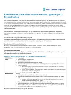
8 Epidemiology Total of 4,563 hospital admissions for burns between 1993 and 1997 o per 100,000 population The Bradford Burn Study (Khan et al 2007) o Studied all burns admissions (n=460) for a full year at a single A&E in the UK o Children of <10 years accounted for 36% of admissions o Wrist and Hand burns accounted for 36% with upper limb burns constituting a further 21% (DORAS 2001) Mechanism of Injury (DOH, Western Australia 2009; Ever et al 2010; Hettiaratchy and Dziewulski 2004) May be thermal or non-thermal 1. Flame burns 50% 2. Scalds from hot liquids, boiling water, cooking oil 40% 3. Contact burn, stoves, heaters, irons, 4. Electrical burn, electrocution 5.
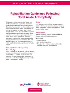
9 Chemical Burns, Hydrofluoric Acid 6. Friction burn 7. Radiation burn Figure 1. Percentage of burn incidents versus age. (Khan et al 2007) 4 Review of the Skin (Tortora and Derrickson 2011) Cutaneous membrane which covers the surface of the body Largest organ of the body in terms of weight and surface area Epidermis o Superficial layer o Composed of epithelial tissue o Avascular o Deepest layer (Stratum Basale) contains Stem cells Capable of regeneration New skin cannot regenerate if injury destroys a large portion of this layer Dermis o Deeper, thicker layer o Connective tissue o Contains blood vessels, nerves, glands and hair follicles Subcutaneous layer o Areolar and adipose tissue o Storage for fat/ insulation o Contains large blood vessels o Attaches to underlying facia Connective tissue overlying muscle and bone Figure 2.
10 Layers of the skin (MD 2009) 5 Table 1 Types of Burns Glassey 2004 Table 1 types of burns (Glassey 2004) Table 1 types of burns (Glassey 2004) Types of Burns 6 Physiology of Burns An in depth knowledge of pathophysiology of burns, and their effects both locally and systemically is necessary to ensure effective management of a patient with a burn injury. Zones of Injury and Wound Conversion The local effect involves three burn zones: (Hettiaratchy and Dziewulski 2004) Zone of Coagulation: the point of maximum damage Irreversible tissue loss due to coagulation of constituent proteins. Zone of Stasis: Characterised by decreased tissue perfusion Potential to rescue the tissue in this zone Problems such as prolonged hypotension, infection or oedema can convert this area into one of complete tissue loss Zone of Hyperaemia: The tissue here will invariably recover unless there is severe sepsis or prolonged hypoperfusion.