Transcription of Prognostic Scoring System for Primary CNS …
1 Prognostic Scoring System for Primary CNS Lymphomas: The international extranodal lymphoma study group Experience By Andre s Ferreri, Jean-Yves Blay, Michele Reni, Felice Pasini, Michele Spina, Achille Ambrosetti, Antonello Calderoni, Andrea Rossi, Vittorio Vavassori, Annarita Conconi, Liliana Devizzi, Franc oise Berger, Maurilio Ponzoni, Bettina Borisch, Marianne Tinguely, Michele Cerati, Mario Milani, Enrico Orvieto, Juvenal Sanchez, Christine Chevreau, Stefania Dell'Oro, Emanuele Zucca, and Franco Cavalli Purpose: To identify survival predictors and to design a patients for which complete data of all five variables were Prognostic score useful for distinguishing risk groups in immu- available. The 2-year overall survival (OS) SD was 80% . nocompetent patients with Primary CNS lymphomas (PCNSL). 8%, 48% 7%, and 15% 7% (P .00001) for patients with Patients and Methods: The Prognostic role of patient-, zero to one, two to three, and four to five unfavorable fea- lymphoma -, and treatment-related variables was analyzed tures, respectively.
2 The Prognostic role of this score was con- in a multicenter series of 378 PCNSL patients treated at 23 firmed by limiting analysis to assessable patients treated with cancer centers from five different countries. high-dose methotrexate-based chemotherapy (2-year OS . Results: Age more than 60 years, performance status SD: 85% 8%, 57% 8%, and 24% 11%; P .0004). (PS) more than 1, elevated lactate dehydrogenase (LDH) Conclusion: Age, PS, LDH serum level, CSF protein con- serum level, high CSF protein concentration, and involve- centration, and involvement of deep structures of the brain ment of deep regions of the brain (periventricular regions, were independent predictors of survival. A Prognostic score basal ganglia, brainstem, and/or cerebellum) were signifi- including these five parameters seems advisable in distin- cantly and independently associated with a worse survival. guishing different risk groups in PCNSL patients.
3 The pro- These five variables were used to design a Prognostic score. posed score and its relevance in therapeutic decision de- Each variable was assigned a value of either 0, if favorable, serve to be validated in further studies. or 1, if unfavorable. The values were then added together to J Clin Oncol 21:266-272. 2003 by American arrive at a final score, which was tested in 105 assessable Society of Clinical Oncology. N SERIES investigating the management of Primary CNS the real efficacy of therapeutic Indeed, apart from age I lymphomas (PCNSL), the differences in outcome observed among various treatment options have been attributed to an and performance status (PS), which are universally accepted Prognostic factors,1,2 no other parameters influencing outcome inhomogeneous distribution of Prognostic indicators rather than have been consistently identified. Efforts to identify predictors of response and survival in PCNSL have produced isolated observations in small series, which have not been confirmed in successive studies.
4 The From the Departments of Radiochemotherapy and Pathology, San Raffaele H characterization of predictors of response and survival using Scientific Institute, and Divisione di Oncologia Medica, Istituto Nazionale dei large series may allow us to identify different patient risk groups, Tumori, Milan; Divisione di Radioterapia, Ospedale di Circolo e Fondazione facilitate the comparative analysis of prospective trials, and Macchi, and Anatomia e Istologia Patologica, Ospedale di Circolo Fondazione define stratification criteria for future trials. In this study , Macchi, Universita` dell'Insubria, Varese; Divisione Clinicizzata di Oncologia predictors of response and survival were analyzed in an interna- Medica, Ospedale Civile Maggiore, and Cattedra di Ematologia, Universita` di Verona, Ospedale Policlinico di Borgo Roma, Verona; Division of Medical tional multicenter retrospective series of 378 immunocompetent Oncology A, Centro di Riferimento Oncologico, National Cancer Institute, patients with PCNSL.
5 A Prognostic score resulting from the Aviano; Divisione di Ematologia, Ospedali Riuniti di Bergamo, Bergamo; combined analysis of the independent variables is proposed in Divisione di Anatomia Patologica, Azienda Ospedaliera di Lecco, Lecco; light of its potential clinical relevance. Istituto di Anatomia e Istologia Patologica, Ospedale Regionale di Treviso, Treviso, Italy; Unite Cytokines et Cancers, Hopital E. Herriot & Centre Le on PATIENTS AND METHODS. Be rard, and Service de Pathologie, Centre Hospitalier Lyon-Sud, Lyon; Centre Claudius Regaud, Toulouse, France; Istituto Oncologico della Svizzera Italiana, study group Ospedale San Giovannia, Bellinzona; Institut fu r Medizinische Onkologie Inselspital, Bern; Division de Pathologie Clinique, Ho pitaux Universitaires de A questionnaire requesting information about patient characteristics, Gene`ve, Ho pital Cantonal, Gene`ve, Switzerland; and Departamento de Anato- clinical presentation, diagnosis, staging, planned and actually performed mia Patolo gica, Instituto de Enfermedades Neopla sicas, Lima, Peru.
6 Treatment, objective response, site and date of relapse, second-line treatment, Submitted September 27, 2001; accepted September 24, 2002. neurotoxicity, and survival was sent to 48 centers referring to the Interna- Presented at the Forty-Second Annual Meeting of the American Society of tional extranodal lymphoma study group . Report forms were submitted to Hematology, December 6-10, San Francisco, CA, 2000 at least one clinician (hematologist or oncologist) and one pathologist per Address reprint requests to Andre s Ferreri, MD, Department of center. Only patient cases diagnosed and treated at the participating institu- Radiochemotherapy, San Raffaele H Scientific Institute, via Olgettina 60, tions that fulfilled the following criteria were selected: (1) histologic or 20132, Milan, Italy; email: cytologic diagnosis of lymphoma ; (2) disease localized exclusively in the 2003 by American Society of Clinical Oncology.
7 Brain, cranial nerves, meninges, or eyes; and (3) no evidence of human 0732-183X/03/2102-266/$ immunodeficiency virus-1 infection (negative serologic tests and absence of 266 Journal of Clinical Oncology, Vol 21, No 2 (January 15), 2003: pp 266-272. DOI: Prognostic FACTORS IN PCNSL 267. epidemiologic risk, opportunistic infections, or lymphopenia for patients Table 1. Patient Characteristics and Extension of Disease diagnosed in the early 1980s) or other immunodeficiencies. The history of a No. of prior cancer was not an exclusion criterion. Patients Thirty-four out of 48 centers responded (response rate, 71%); 11 centers Characteristic (N 378) %. did not register patients in the study . Therefore, data of 378 PCNSL patients Age, years diagnosed between 1980 and 1999 were collected by 23 cancer centers (48% Median 61. of responding centers) from five different countries. Range 14-85. Questionnaire data were verified during a consensus meeting held at Age 70 years 52 14.
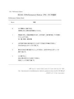
8 Ascona, Switzerland, on February 2000; equivocal submitted information Male sex 220 58. was subsequently analyzed until consensus for every single patient case was ECOG PS. reached. Each institution carried out a radiologic material review. Clinical 0-1 117 31. staging work-up included at least total-body computed tomography scan and 2-3 175 46. bone marrow biopsy. Staging procedures included slit-lamp examination of 4 47 12. the eyes in 170 patients (45%) and CSF cytology examination in 241 patients Not specified 39 10. (64%). The cutoff for normal CSF protein concentration was 45 mg/dL in Prior cancer 16 4. patients 60 years old and 60 mg/dL in patients more than 60 years Histotype*. PS was defined according to the Eastern Cooperative Oncology group A-C 11 3. (ECOG) criteria. Histologic sample was obtained by stereotactic biopsy in D-G 220 58. 276 patients (72%), surgical resection in 86 patients (23%; total resection in H-K 77 20.)
9 44 patients, subtotal in 42 patients), CSF cytology examination in 14 patients Unclassified 70 19. (4%), and vitrectomy and autopsy in one patient each ( 1%). T cell 8 2. Systemic symptoms 7 2. Therapeutic Management LDH ratio 1 69/195 35. Therapeutic data were available from 370 patients. Planned treatment was Ocular disease 22/170 13. radiotherapy (RT) alone in 98 patients (26%), RT followed by chemotherapy Positive CSF cytology examination 38/241 16. (CHT; RT 3 CHT) in 36 patients (10%), CHT alone in 32 patients (9%), and High CSF protein level 82/134 61. CHT followed by RT (CHT 3 RT) in 197 patients (53%). Seven patients Multiple lesions 130 34. (2%) did not receive any treatment. It is noteworthy that five patients who Involvement of deep structures 136 36. were planned to receive Primary RT did not receive any treatment, whereas Site of disease 61 patients who were planned to receive CHT 3 RT were not irradiated Frontal lobe 166 44.
10 Because of progressive disease (n 25), early relapse (n 11), toxic death Parietal lobe 50 13. (n 15), refusal (n 6), unrelated death (n 1), or because they were lost Temporal lobe 52 14. to follow-up (n 3). Occipital lobe 24 6. Methotrexate (MTX) was the most commonly used drug (n 169), Basal ganglia 104 28. followed by alkylating agents (n 146, including procarbazine, thiotepa, Brain stem 21 6. cyclophosphamide, and nitrosoureas), high-dose (HD) cytarabine (n 128), Cerebellum 23 6. and anthracyclines (n 117). MTX was administered at a dose 3 g/m2 per Meninges 39 10. course in 126 patients (75%). CHT regimen was alkylating agents alone in 25 Cranial nerves 3 1. patients, cyclophosphamide, doxorubicin, vincristine, and prednisone *Histotype was defined according to the Working Formulation Classification. (CHOP) or CHOP-like regimens in 43 patients, HD cytarabine-based Relationship between the number of positive cases and the total number of regimen in 11 patients, HD-MTX alone in 17 patients, HD-MTX plus assessed patients.