Transcription of Progressive Late-Onset Cerebellar Ataxia A - ACNR
1 Review Article Progressive Late-Onset Cerebellar Ataxia A. taxia is a term used to describe a condition charac- suggest there are at least 10,000 cases of familial and spo- terised by disordered or incoordinate movement and radic Late-Onset Cerebellar Ataxia (LOCA) in the UK alone, is commonly caused by diseases affecting the cerebel- with the majority of both familial and sporadic cases hav- lum and its connections within the central nervous system ing no defined aetiology. (CNS). Ataxia caused by Cerebellar dysfunction is a domi- nant feature in a wide spectrum of overlapping heteroge- Diagnostic strategy neous clinical disorders1 and may also be mimicked by a For many patients, especially those with an acute or sub- variety of isolated or combined neurological deficits, includ- acute presentation of Ataxia , initial investigations will read- ing loss of muscular strength, altered tone, diminished sen- ily identify an acquired cause (Table 1) such as chronic Dr Mark Wardle is a Specialist sation or the intrusion of involuntary movements.
2 Alcoholism, MS, remote malignancy (paraneoplastic cere- Registrar in Neurology in Cardiff, Ataxia may present either as a pure Cerebellar syndrome bellar degeneration), vitamin deficiency, toxins or currently undertaking research on or associated with significant cognitive, pyramidal, extra- Adult patients with a more Progressive Cerebellar Ataxia . pyramidal, sensory and autonomic dysfunction, and can disease course of more than one year, particularly if asso- also be the presenting feature of a more widespread neu- ciated with few discriminatory signs on neuroimaging, rodegenerative disorder. It is therefore not surprising that commonly present the most challenging clinical scenario. the investigation of Cerebellar Ataxia often poses consider- They require careful initial and subsequent clinical assess- able diagnostic challenges for the treating physician, and ment of frequently complex clinical features which may has been made increasingly complex by advances in change over time.
3 This necessitates a practical diagnostic molecular genetics and immunology that allow access to a and management strategy, focusing on the early identifica- bewildering array of novel investigations. tion of potentially reversible causes and demands a logical approach to more specialised investigations (Table 2). Extent of the problem The most important discriminating factors in the history Dr Neil Robertson is a Senior Cerebellar Ataxia is not a rare condition: Hospital Episode and examination of this group of patients with late onset Lecturer and Honorary Consul- Statistics (HES) for England and Wales (2005/6) suggest Cerebellar Ataxia (LOCA) are family history, age of onset, tant Neurologist in Cardiff with an admission figures similar to disorders such as myasthenia rate and pattern of development of symptoms, a compre- interest in neuro-inflammatory disease and genetic epidemiology.
4 Gravis, idiopathic intracranial hypertension and bacterial hensive drug and alcohol history, past medical history and meningitis, and three times that of Huntington's disease the presence of other associated symptoms and signs. (HD) ( ). However, these statis- Correspondence to: tics are likely to significantly underestimate the true extent Is there a family history? Dr Mark Wardle and Dr Neil Robertson, of the problem as Cerebellar disease frequently occurs as a A dominant family history is the single most important fac- Department of Neurology, feature of other primary neurological disorders such as tor predicting the chance of successfully diagnosing a genet- Ophthalmology and multiple sclerosis (MS), stroke and CNS tumours. Accurate ic cause of Cerebellar Ataxia .
5 Analysis of the common spin- Audiological Medicine, epidemiological statistics for incidence and prevalence are ocerebellar mutations results in a positive identification in School of Medicine, Cardiff University, scarce and highly variable largely as a result of ascertainment 39 64% of dominant and 11-38% of non-dominant fami- Heath Park, bias, differing inclusion criteria, variable aetiological classi- lies, but only 1 19% of sporadic Late-Onset 13 Cardiff, CF14 4XN, UK. fication and founder effects. Contemporary estimates of the The inherited ataxias are a broad heterogeneous group, Email. prevalence of autosomal dominant Cerebellar Ataxia and can manifest in childhood, adolescence or adulthood (ADCA) in the UK lie between ,0002-4 and with widely variable clinical features, even within the ,000 7 same family.
6 They may be inherited in an autosomal Prevalence estimates for sporadic idiopathic Late-Onset recessive, autosomal dominant (ADCA), X-linked or Cerebellar Ataxia are limited, but a minimum prevalence of mitochondrial inheritance pattern, but prevalence esti- ,000 has been suggested for the These data mates are limited and sensitive to founder effects resulting Table 1: Aetiology of Cerebellar Ataxia . Adapted from reference 34. Acute (hours to days) Subacute (days to weeks). Intoxication (alcohol, lithium, barbiturates) Intoxication (mercury, solvents, petrol, glue, cytotoxic agents). Acute viral cerebellitis Alcoholic Cerebellar degeneration Post-infection syndrome Nutritional / malabsorption (vitamin B1, vitamin B12). Vascular ( Cerebellar infarction, haemorrhage) Posterior fossa tumour ( Cerebellar glioma, metastases).
7 Infectious ( Cerebellar abscess, Whipple's) Multiple sclerosis Hydrocephalus Chronic (months to years) Foramen magnum compression Intoxication (phenytoin toxicity) AIDS-related multi-focal leukoencephalopathy Paraneoplastic Cerebellar syndrome Miller-Fisher syndrome Gluten Ataxia ' Lyme disease Vitamin E deficiency (inherited or acquired). Hypothyroidism Episodic Ataxia Tabes dorsalis Intoxication Creutzfeld-Jacob disease Multiple sclerosis Rubella panencephalitis Transient ischaemic attacks Previous vascular lesion or demyelination Foramen magnum compression Congenital lesion Intermittent hydrocephalus ( cysticercosis, colloid cyst). Inherited ataxias Dominant episodic Ataxia ( EA1,EA2 etc.). Idiopathic'/degenerative ataxias Inherited metabolic ataxias 6 I ACNR VOLUME 7 NUMBER 2 MAY/JUNE 2007.
8 Review Article lutamine track in the resulting protein leading to abnormal protein confor- mation; others are either untranslated repeats, deletions or missense muta- tions. The five main pathogenetic mechanisms of inherited ataxias are abnormal protein folding ( SCA1), mitochondrial ( Friedreich's Ataxia ), defective DNA repair ( Ataxia telangiectasia), channelopathies ( EA1) and metabolic ( inherited vitamin E deficiency).15. Which genetic tests? NHS laboratories across the UK commonly perform SCA1, SCA2, SCA3, SCA6 and usually SCA7 in response to a generic SCA screen' request. Other investigations such as SCA12, SCA17, DRPLA, and Friedreich's are available but usually must be specifically requested. Research laboratories may offer additional tests (Table 3) if the clinical circumstances are appropriate.
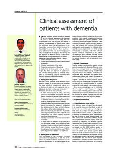
9 The choice of diagnostic tests should be guided by local knowledge of the common Ataxia families, an insight into the prevalence of common SCA subtypes (Figure 1) within the ethnic group, and the presence of suggestive extracerebellar features (Table 4). However, pheno- typic variability and overlap make clinical diagnosis difficult, and in most cases, screening for a range of diseases is necessary. If there is a strong dominant family history, it is appropriate to screen for SCA1, SCA2, SCA3, SCA6 and SCA7 as part of first-line investiga- Figure 1: Aggregated data showing ethnic differences in SCA subtype frequency among tions. An important clue to the presence of a dominantly inherited trin- families with ADCA. ucleotide repeat (TNR) disorder is anticipation, resulting in increasing severity and earlier age of onset through generations.
10 However, even in sporadic cases, a routine screen is recommended since the presence of a in variable frequencies of the different subtypes both geographically and dominant family history may be hidden by reduced penetrance ( ethnically. SCA1, SCA2 and SCA3 are the commonest cause of ADCA in SCA17), marked anticipation (most notable in SCA7) or false paternity. Caucasian families, but SCA3, SCA6 and DRPLA are more common in In sporadic cases, or if there is a history of consanguinity or affected sib- Asian populations (Figure 1). lings, testing for Friedreich's Ataxia (FA) is essential. FA is the most com- A clinical classification of autosomal dominant Cerebellar Ataxia (ADCA) mon recessive cause of spinocerebellar Ataxia , and traditionally this clini- was introduced by Harding in 1982,14 with a division into ADCA I,II and III cal diagnosis was limited to patients with an onset below the age of 25 with based on the presence of extracerebellar features, a pigmentary retinopathy or a pure Cerebellar syndrome respectively.