Transcription of Protein Structure - particlesciences.com
1 L-ArginineArgRL-LysineLysKL-HistidineHis HFigure 1 Particle Sciences QL-GlutamineGlnL-ThreonineThrTL-Methioni neMetMH2 ONL-IsoleucineIleIAsnNL-AsparagineL-Prol ineProPL-TyrosineTyrYL-Aspartic acidAspDL-Glutamic acidGluEGlycineGlyGL-AlanineAlaAL-Trypto phanTrpWL-PhenylalaninePheFCysCL-Cystein eL-LeucineLeuLL-ValineValVL-SerineSerSOH 2NH2 NBasicAcidicNeutralOOOHH2NH2 NOOHOOOHNH2H NH2 NNHHNNHNH2 ONH2 ONH2 OHONH2 OHONOHH2 ONH2 ONH2 ONH2 ONH2 SHONH2 OHOHOHOHOHOHOHOHOHOHOHOHOHOHOHOHOHOHOHON H2 ONHONH2 NOSOOH2NH2 NNH2 OOH2 NProtein StructureTechnical Brief 2009 Volume 8 Increasingly, drug developers are looking to large molecules and partic-ularly proteins as a therapeutic option. Formulation of a Protein drug product can be quite a challenge, but without a good understanding of the nature of Protein Structure and the conforma-tional characteristics of the specific Protein being formulated, the results can be ruinous. This technical brief aims to give the reader a quick over-view of Protein Structure .
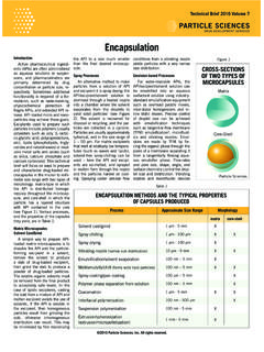
2 It will also cover briefly how Protein Structure can be affected during formulation and some of the analytical methods which can be used both to determine the Structure and analyze the stability of the Protein . The term Structure when used in relation to proteins, takes on a much more complex meaning than it does for small molecules. Proteins are mac-romolecules and have four different levels of Structure primary, second-ary, tertiary and quaternary. Primary StructureThere are 20 different standard L- -amino acids used by cells for Protein construction. Amino acids, as their name indicates, contain both a basic amino group and an acidic car-boxyl group. This difunctionality al-lows the individual amino acids to join together in long chains by forming peptide bonds: amide bonds between the -NH2 of one amino acid and the -COOH of another. Sequences with fewer than 50 amino acids are gener-ally referred to as peptides, while the terms Protein or polypeptide are used for longer sequences.
3 A Protein can be made up of one or more polypeptide molecules. The end of the peptide or Protein sequence with a free carboxyl group is called the carboxy-terminus or C-terminus. The terms amino-ter-minus or N-terminus describe the end of the sequence with a free -amino group. The amino acids differ in struc-ture by the substituent on their side chains. These side chains confer dif-ferent chemical, physical and struc-tural properties to the final peptide or Protein . The structures of the 20 ami-no acids commonly found in proteins are shown in Figure 1. Each amino acid has both a one-letter and three-letter abbreviation. These abbrevia-tions are commonly used to simplify the written sequence of a peptide or Protein . Depending on the side-chain sub-stituent, an amino acid can be classi-fied as being acidic, basic or neutral. Although 20 amino acids are required for synthesis of various proteins found in humans, we can synthesize only 10.
4 The remaining 10 are called es-sential amino acids and must be ob-tained in the diet. The amino acid sequence of a pro-tein is encoded in DNA. Proteins are synthesized by a series of steps called transcription (the use of a DNA strand to make a complimentary messenger RNA strand - mRNA) and translation (the mRNA sequence is used as a template to guide the synthesis of the chain of amino acids which make up the Protein ). Often, post-translational modifications, such as glycosylation or phosphorylation, occur which are necessary for the biological func-tion of the Protein . While the amino acid sequence makes up the primary Structure of the Protein , the chemical/biological properties of the Protein are very much dependent on the three-di-mensional or tertiary StructureStretches or strands of proteins or peptides have distinct characteris-tic local structural conformations or secondary Structure , dependent on hy-drogen bonding.
5 The two main types of secondary Structure are the -helix and the -sheet. The -helix is a right-handed coiled strand. The side-chain substituents of the amino acid groups in an -helix extend to the outside. Hydrogen bonds form between the oxygen of the C=O of each peptide bond in the strand and the hydrogen of the N-H group of the peptide bond four amino acids below it in the helix. The hydro-gen bonds make this Structure espe-cially stable. The side-chain substitu-ents of the amino acids fit in beside the N-H groups. The hydrogen bonding in a -sheet is between strands (inter-strand) rath-er than within strands (intra-strand). The sheet conformation consists of pairs of strands lying side-by-side. The carbonyl oxygens in one strand hydrogen bond with the amino hy-drogens of the adjacent strand. The two strands can be either parallel or anti-parallel depending on whether the strand directions (N-terminus to C-terminus) are the same or oppo-site.
6 The anti-parallel -sheet is more stable due to the more well-aligned hydrogen bonds. Tertiary StructureThe overall three-dimensional shape of an entire Protein molecule is the tertiary Structure . The Protein mol-ecule will bend and twist in such a way as to achieve maximum stability or lowest energy state. Although the three-dimensional shape of a Protein may seem irregular and random, it is fashioned by many stabilizing forces due to bonding interactions between the side-chain groups of the amino acids. Under physiologic conditions, the hydrophobic side-chains of neutral, non-polar amino acids such as phe-nylalanine or isoleucine tend to be buried on the interior of the Protein molecule thereby shielding them from the aqueous medium. The alkyl groups of alanine, valine, leucine and isoleucine often form hydrophobic interactions between one-another, while aromatic groups such as those of phenylalanine and tryosine often stack together.
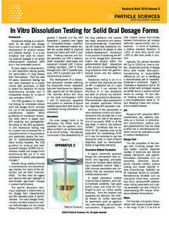
7 Acidic or basic amino acid side-chains will generally be ex-posed on the surface of the Protein as they are hydrophilic. The formation of disulfide bridges by oxidation of the sulfhydryl groups on cysteine is an important aspect of the stabilization of Protein tertiary Structure , allowing different parts of the Protein chain to be held together covalently. Additionally, hydrogen bonds may form between different side-chain groups. As with disulfide bridges, these hydrogen bonds can bring together two parts of a chain that are some distance away in terms of sequence. Salt bridges, ionic inter-actions between positively and nega-AMINO ACID STRUCTURES AND ABBREVIATIONSP article Sciences Figure 2 HHNOHOCNCHCONNNNNNNNCHHHHHHHHCOCCCCCCCCO COCOCOCOCOCONNHCCOCOCONHCNNHCCCOCONHHCCC COCOCONHNNHHCCCCOCONHH3894 Courtney StreetBethlehem, PA 18017-8920, USAP hone: +1 610 861 4701 Fax: + 610 861 4702 Email: Sciences is a leading integrated provider of formulation and analytic services and both standard and nanotechnology approaches to drug development and charged sites on amino acid side chains, also help to stabilize the tertiary Structure of a StructureMany proteins are made up of multiple polypeptide chains, often referred to as Protein subunits.
8 These subunits may be the same (as in a homodimer) or different (as in a het-erodimer). The quaternary Structure refers to how these Protein subunits interact with each other and arrange themselves to form a larger aggregate Protein complex. The final shape of the Protein complex is once again stabilized by various interactions, in-cluding hydrogen-bonding, disulfide-bridges and salt bridges. The four levels of Protein Structure are shown in Figure StabilityDue to the nature of the weak in-teractions controlling the three-di-mensional Structure , proteins are very sensitive molecules. The term native state is used to describe the Protein in its most stable natural conformation in situ. This native state can be dis-rupted by a number of external stress factors including temperature, pH, re-moval of water, presence of hydropho-bic surfaces, presence of metal ions and high shear. The loss of secondary, tertiary or quaternary Structure due to exposure to a stress factor is called denaturation.
9 Denaturation results in unfolding of the Protein into a random or misfolded shape. A denatured Protein can have quite a different activity profile than the Protein in its native form, usually los-ing biological function. In addition to becoming denatured, proteins can also form aggregates under certain stress conditions. Aggregates are of-ten produced during the manufactur-ing process and are typically undesir-able, largely due to the possibility of them causing adverse immune re-sponses when administered. In addition to these physical forms of Protein degradation, it is also im-portant to be aware of the possible pathways of Protein chemical deg-radation. These include oxidation, deamidation, peptide-bond hydroly-sis, disulfide-bond reshuffling and cross-linking. The methods used in the processing and the formulation of proteins, including any lyophilization step, must be carefully examined to prevent degradation and to increase the stability of the Protein biophar-maceutical both in storage and during drug delivery.
10 Protein Structure AnalysisThe complexities of Protein struc-ture make the elucidation of a com-plete Protein Structure extremely dif-ficult even with the most advanced analytical equipment. An amino acid analyzer can be used to deter-mine which amino acids are present and the molar ratios of each. The sequence of the Protein can then be analyzed by means of peptide map-ping and the use of Edman degrada-tion or mass spectroscopy. This pro-cess is routine for peptides and small proteins, but becomes more complex for large multimeric mapping generally entails treatment of the Protein with different protease enzymes in order to chop up the sequence into smaller peptides at specific cleavage sites. Two com-monly used enzymes are trypsin and chymotrypsin. Mass spectroscopy has become an invaluable tool for the analysis of enzyme digested proteins, by means of peptide fingerprinting methods and database searching.