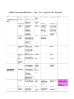
Transcription of Pulmonary Function Tests - Columbia University
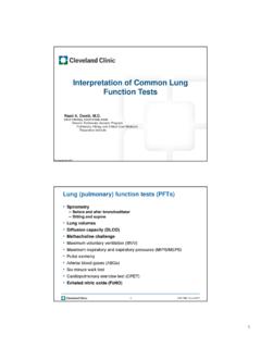
1 Pulmonary Function Tests PFT Interpretation The interpretation of lung Function Tests involves two tasks: 1) the classification of the derived values with respect to a reference population and assessment of the reliability of the data; and 2) the integration of the obtained values into the diagnosis, therapy and prognosis for an individual patient. ATS/ERS TASK FORCE: STANDARDISATION OF LUNG Function TESTING''. Eur Respir J 2005; 26: 153 161. Pulmonary Function : Tests Dynamic Function : obstructive defects Static Function : restrictive defects Diffusion abnormalities (gas exchange). Spirometry and Maximal Expiratory and Spirometry Inspiratory Flow Volume Curves Dynamic Function . 1. Obstructive Ventilation: Expiratory Decrease in expiratory airflow (volume and/or rate of flow). FEV1 decreased FVC normal or decreased FEV1/FVC decreased*.
2 FEF25-75 decreased *definition of obstructive defect Types of Airflow Obstruction Bronchoconstriction Dynamic airway compression (FVC vs SVC). Emphysema: FVC < slow or inspiratory VC, and plethysmographic volumes greater than gas dilution volumes Upper Airway Small Airways Mixed . 2. PFT Question #1. FEV1 /FVC=obstructive ventilatory defect: Why is FEV1 itself NOT diagnostic of an obstructive defect? Upper Airway Obstruction 3. Upper Airway Obstruction Lung Volumes Static Function . Gas Equilibration ( wash in and wash out ). Body plethysmography Gas Equilibration Lung Volumes Wash in: Helium (insoluble gas) breathed from a reservoir of known VOLUME and CONCENTRATION, thus diluting its concentration by the volume of the lungs VFRC = Vreservoir x Conc INIT Conc FINAL/ Conc FINAL. Gas Equilibration Lung Volumes Plethysmographic Lung Volumes Wash out: Lung gas (N2) washed out P1V1=P2V2 in a closed system at same during breathing of 100% O2 temperature Initial N2 concentration known Lungs and airway closed system when (atmospheric); volume and N2 occluded concentration of expired gas measured Panting at FRC: inhalation=decreased VFRC=VEXP X conc EXP/.
3 79- Conc ALV (final) intrathoracic pressure, increased volume 4. Plethysmographic Lung Volumes VFRC=V / P (PFRC- P ) where P is negligible c/w PFRC. VFRC= V / P (PFRC). P obtained from change in mouth pressure against occluded valve V obtained from change in pressure in the plethysmograph as air in the box is compressed by increase in lung volume PFT Question #2. Measurement of Alveolar In airways disease ( , emphysema), if gas dilution is not complete, how will lung Volume (VA). volume measurement be affected? VA pleth > VA He rebreathe > >VA He single breath VA He rebreathe > VA single breath correlated with decreased FEV1/FVC, increased RV/TLC. Restrictive Ventilation PFT Questions #3 and #4. A decrease in lung expansion Why is FVC itself NOT diagnostic of a FEV1 decreased restrictive ventilatory defect? FVC decreased Why is VC itself not diagnostic of a FEV1/FVC normal or increased restrictive ventilatory defect?
4 Total Lung Capacity (TLC) decreased*. * Definition of restrictive ventilatory defect 5. Types of Restrictive Defects Restrictive patterns Parenchymal removal/destruction Diffuse parenchymal disease, thoracic cage Parenchymal infiltration restriction: symmetric decrease in TLC, Extrapulmonary deformity VC, FRC, RV. Neuromuscular weakness: IC mainly Reduced force generation decreased; TLC and VC decreased and FRC and RV spared Diffusing Capacity (Transfer Factor). Diffusing Capacity for CO (DLCO) Diffusing Capacity for CO (DLCO). DLCO = CO rate of uptake (ml/min)/ PCO DLCO (if transfer factor, TLCO) calculated as the product of (mmHg) the rate constant for CO uptake (called kCO, the Krogh O2 and CO combine with Hgb; therefore reflect coefficient) and alveolar volume, divided by effective gas properties of alveolar-capillary membrane, and its pressure (PB-PH20), expressed as units of conductance uptake therefore limited by resistance across this (eg, ml CO/min/mmHg).
5 Interface Soluble gases limited by Pulmonary blood flow Thus, DLCO =(kcOxVA)/(Pb-PH20). 2 major resistances therefore: membrane properties (Dm), and reactive conductance This assumes what the conductance would be if 100% of (molecular conformation/rate of reaction alveolar volume was filled with CO (that is the VA component properties of Hgb binding x Pulmonary capillary is the volume of distribution). blood volume (Vc).. 6. SB Diffusing Capacity for CO. Diffusing Capacity for CO (DLCO). (DLCO) Inspirate CO, 10% inert gas, 21%O2, balance N2. Expire to RV; inhale rapidly to TLC; hold for remainder of 10 seconds of breath hold time (BHT). Diffusion determinants: Gas gradient, Expire; discard anatomic dead space gas;. solubility, hemoglobin, membrane sample 500-1000 ml alveolar gas thickness, surface area Diffusing Capacity Increased in alveolar hemorrhage, obesity, asthma?)
6 ?, altitude (since CO and O2 in competition, altitude decreases PIO2 and increases DLCO). Decreased in emphysema (destruction and/or non- equilibration), restrictive disorders (all:why??), Pulmonary vascular disorders, anemia, abnormal Hgb Single breath (10 sec) vs steady state/rebreathe techniques: SB may UNDERESTIMATE true diffusing capacity in emphysema if it underestimates gas dilution VA since DLCO. =(kcOxVA)/(Pb-PH20. DLCO Pearl Isolated DLCO decrease: suspect Pulmonary vascular disorder Or, interstitial disorder not yet, or no longer, affecting parenchymal volume Or, abnormality of Hgb (eg, anemia, carboxyhgb, methhgb). 7. Pre-operative Pulmonary Pre-operative Pulmonary Assessment: PFTs Assessment: PFTs Complications: highest for thoracic and Spirometry: FEV1 or FVC <70%, upper abdominal (ie, near the diaphragm) FEV1/FVC<65%.)
7 All having lung resection, orthopoedic and PaCO2>45 mmHg, DLCO<40% in COPD. lower abdominal with lung disease, or None contraindicate smoking Lung resection: FEV1 best for Pulmonary Age>60 years reserve and post op complications; post op FEV1 <30% predicted=increased long term mortality and immediate post op problems PFT Summary Series ATS/ERS TASK FORCE: Obstructive ventilatory defect: decreased STANDARDISATION OF LUNG. FEV1/FVC. Restrictive ventilatory defect: decreased TLC. Function TESTING'' Edited by Low DLCO: abnormal uptake of gas by Hgb V. Brusasco, R. Crapo and G. across alveolar capillary membrane: Diffusion Viegi. General considerations for determinants= Gas gradient, solubility, hemoglobin, membrane thickness, surface area lung Function testing Disorders with airway dysequilibration (emphysema): gas dilution will underestimate Eur Respir J 2005; 26: 153 161.
8 Lung volumes (and ? DLCO). When you can't breathe, nothing else matters.. 8.