Transcription of Radiation safety in dental radiography
1 dental radiography SeriesRadiation safety indental radiographyThe goal of dental radiography is to obtain diagnosticinformation while keeping the exposure to the patient anddental staff at minimum some exposure to Radiation is acceptable in medicalpractice, it should be understood that levels of radiationexposure to patients, dental staff, and other nearbyoccupants should be kept to As Low As ReasonablyAchievable (ALARA) to reduce health risks from methods that can reduce patient and area radiationexposures without major difficulty, great expense orinconvenience, should be practiced.
2 Practitioners mustalways consider the risk of patient exposure with the benefitof guidelinesOne way to do this is with the use of radiographic patient selection for the prescription of dental radiographshave been developed by an expert panel of dentistssponsored by the public health free brochure is available from Carestream Health,Inc. (see last page for ordering information) publication N-80A Guidelines for prescribing dental radiographs. The guide-lines are voluntary and are intended only as a decision-making aid for the dentalpractitioner.
3 They are used only in conjunction with a carefully taken medical anddental history and a clinical safety considerationsIn any case, once the decision has been made to prescribex-rays, every reasonable effort must be made to minimizeexposure to the patient and dental office , the same safety procedures that minimizeexposure for both patient and operator can also increasethe quality of the radiographic are many factors that determine the level ofradiation received by the patient during a radiographicexamination.
4 These include: The selection of the x-ray machine The use of technique factors that result in low patient exposure The use of fast films and screen/film combinations Adherence to correct film processing methods The use of digital sensors The use of collimators and filtration The use of lead aprons and thyroid collars to protect the patient fromunnecessary Radiation exposureAll x-ray equipment, regardless of date of manufacture, is subject to state andfederal x-ray equipment proper filtration is not usually a problem with modern equipment, olderx-ray machines should be tested by a Radiation physicist or qualified technicianto verify the presence of the correct amount of kilovoltage or kVp setting is one of the most important factors thatdetermines the image contrast, as well as dosage to the patient.
5 In the 60-80 kVp range, biological risk esti mates from dental radiology are essentiallythe same and, therefore, the diagnostic need should be the determining factorfor which kVp setting to use. Settings below 60 kVp are not recommended forroutine dental radiography because of higher patient Radiation skin entry exposures and effective dose equivalents are outlined in the tablebelow:Typical patient doses from dental x-ray examsTo keep the effective dose equivalent in perspective; in 1991, a research team at theAcademic Center for Dentistry in Amsterdam made an elaborate series of measurements ofdose to all areas of the head and neck during bite-wing radiography using a plastic "headphantom.
6 " They found an effective dose for the bite-wing of mrem. To put this inperspective, background Radiation from naturally occurring radionuclides in our environmentand from cosmic rays delivers almost a thousand times as much Radiation every year(approximately 300 mrem).The benefits of the use of x-rays in dentistry outweigh the risks when proper safetyprocedures and considerations are dentist or registrant of the x-ray producing equipment is responsible for all aspects ofradiation safety in the dental dentist selects the patient who needs radiographs,determines which radiographs are needed.
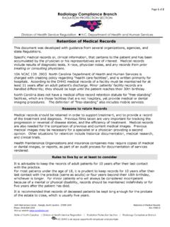
7 Takes or supervisesthe exposure of the films and interprets the important method for keeping patient exposure as low asreasonably achievable is the appropriate prescription mouth (18 exposures)2,300-3, Dose (mR)Effective Dose (mrem)3 Rectangular collimationCollimators, when installed properly, serve tolimit the size and shape of the useful x-ray beamreaching the patient. This will not only reducedose, but may also improve image American dental Association ( ) andthe American Academy of Oral and MaxillofacialRadiology recommend the use of a shielded,open-ended, position-indicating device, or PID,preferably with rectangular is an example of a rectangular collimatorthat restricts the beam to the size and shape ofthe dental film.
8 Round collimators can be converted to a rectangular shapedopening by using an insert avail able through a manufacturer of dentalradiographic products (see last page for suggested resource). This techniquesignificantly reduces the volume of tissue exposed during intraoral collimators reduce patientexposure by restricting the beam size tothat of the film used. These devices willincrease subject contrast by reducingexcessive scatter area and volume of tissue exposed tothe primary x-ray beam should not exceedthe minimum coverage required to imagethe anatomical area in question.
9 Periapicalradiographs should, in general, demonstrate 1/4-in of alveolar bone beyond theapex of each tooth, 1/8- to 1/4-in margin between the crowns of the teeth andthe edge of the film; the occlusal plane should be straight or slightly curvedupward toward the bite-wing views, the occlusal plane should be straight or slightly curvedupward toward the distal. There should be equal distribution of maxillary andmandibular crowns and maxillary and mandibular alveolus, and the interproximalspaces should be open.
10 These criteria can be met successfully by carefulexecution of correct periapical and bite-wing holding devices are recommended for intraoral radiography toeliminate the need for the film to be held in place by the patient sfinger. These film holding devices also provide for the film to beplaced parallel to the teeth, resulting in a less distorted image. Theholders recommended today incorporate beam guiding deviceswhich make PID alignment a simpler and the Academy discourage the use of short, closed,pointed plastic cones because of the increased scatter radiationand unnecessary Radiation close to the face and surrounding areasof the patient.