Transcription of Rare Cranio-Facial clefts - Cirujanos Plastikos Mundi
1 Rare Cranio-Facial clefts Plastic and Reconstructive Surgery Cirujanos Pl stiKos Mundi 1. Rare Cranio-Facial clefts 1. Introduction. Cranio-Facial clefts are a wide spectrum of malformations affecting the face and cranium in a great variety of forms. The low frequency of most of them has made its study, statistics and classification complex and uncertain for many, many years. clefts in the Cranio-Facial region range from the most commonly known cleft lip and palate to extensive Cranio-Facial clefts that can be dramatically disfiguring. facial clefts constitute the most challenging malformation as they are never the same. The surgeon must be skilful in Cranio-Facial surgery, maxillofacial techniques, soft tissues procedures for soft tissue reconstruction, and no less important, have a solid background in management of Cranio-Facial anomalies. Despite all this, restoring, functional and aesthetically, a clefted face is one of the most rewarding surgeries.
2 Brief history of classification efforts to bring order out of chaos (in words of Henry Kawamoto), description of the clefts and principles for surgery will be discussed in this chapter. 2. Classification of Cranio-Facial clefts . Cranio-Facial malformations, and between them, facial clefts , were first describe by doctors, anathomist, genetist, etc, etc. The general rule was to nominate each malformation with its own name, been the case of the same or similar malformation known with various names at the same time (Sdme of Franceschetti, Goldenhar, 1st and 2nd Brachyal sdmes., Treacher-Collins, Apert, Crouzon, etc, etc). After interest for this pathology has grown, several attempts to classified them had been carried out. Morian Classification Morian, in 1887, was the first to be credited for presenting a classification for facial clefts . He described two types, in which the reference was the infraorbital foramen.
3 2. Type I, the oculonasal clefts , in which clefts occupied the region between the infraorbitary foramen and the middle line of the face, and, Type II, from the infraorbital foramen to the outer aspect of the face. AACPR Classification In 1962, the American Association of Cleft Palate Rehabilitation (AACPR), described a new classification for facial clefts and syndromes and divided them in four major groups : 1st. Group : Mandibular process clefts . 2nd Group : Naso-ocular clefts . 3rd Group : Oro-ocular clefts . 4th Group : Oro-auricular clefts . This classification does not include major midline facial clefts , Treacher-Collins Syndrome and does not integrate the underlying bone defects. So it was an incomplete classification. Boo-Chai Classification The deficiencies of previous classifications were clear for Boo-Chai, and based on the classification already described by Morian, amplified the description of oro-ocular clefts , by subdividing them into types I or II.
4 Karfik Classification In 1966 Karfik was to consider by first time embryological and morphological aspects of the Cranio-Facial deformities. He described five groups : A group : Rhinencephalic disorders B group : 1st. and 2nd arch brachial disorders C group : Ophthalmo-orbital disorders D group : Cranio-cephalic disorders E group : Atypical facial disorders Other classifications were made various authors as Lund, DeMeyer, etc., but the leading of all was made by Paul Tessier, he presented it in 1973 during the 1st international congress of Cleft Palate, and was later popularized by Kawamoto in 1976. Tessier Classification This classification is the generally accepted one for description of all the cranio- facial clefts . It represents an orderly anatomic classification system in which all the clefts , major and minor and despite its position are considered and numbered from 0 to 14 with a number 30 for a medial symphysis in the mandible.
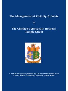
5 This system significantly simplify the nomenclature of clefts . It is a purely descriptive system not related to embryological or pathological factors. 3. Fig. 1 a) and b) Tessier Classification on the skull and face The system followed by Tessier is centred on the orbit, with facial clefts numbered from 0 to 7 and the cranial clefts from 8 to 14 in a counter clockwise rotation, being the 30 in the midline of the mandibular symphysis. The orbit is the central point of the schema and separates the facial structure from the cranium. Other attempts had been made posteriorly by others groups as that of Van de Meulen in which correlation between the clefts and embryological events are the basis. However Tessier classification is easiest for its way of description and the simplicity of its nomenclature, and remains the more practical classification until now. The clefts may involve soft tissues and bony structures in a different degree at different levels and can occur independently.
6 clefts could be presented unilaterally or bilaterally, though unilateral forms are the most common. clefts tend to follow the same axis, so is important to explore them very carefully in order to diagnose other features, though it does not mean that a cleft affecting soft tissues in the surface will affect in the same manner the underlying structures. Even more,defects on the bony structures are greater in clefts affecting the face than those in the cranium. The extent and disfigurement caused by a cleft varies considerably, so a cleft could be as complete as to affect all the soft tissues with all the underlying bony structures or just be manifested as simple skin notch or small bridle in the oral mucosa. 3. Etiology 4. Formation of clefts takes place while the embryo is growing, and there is a pattern for the basic types of different clefts , so is important to know something about the embryogenesis of the face as a principle to understand these complex malformation.
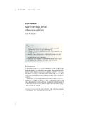
7 Fig. Frontal process Lateral nasal part of frontal process Medial nasal part of frontal process Maxilary process Mandibulary process Frontal process Lateral nasal part of frontal process Medial nasal part of frontal process Maxillary and Mandibular processes The different processes will joint together around the mouth, so anything that would interfere this meeting place or would cause the union to be ruptured will provoke a cleft. Correlation between the processes in the face of an embryo and the face of an adult will help to understand the morphology and distribution of clefts regarding the mechanics and chronology of Cranio-Facial morphogenesis. 5. Fig. Correlation between the processes of the face of an embryo and an adult Etiology of Cranio-Facial clefts is based on the same theories and principles as those described for cleft lip and palate. In fact, is the research on these more common clefts what has open the ways for the two main theories, that of failure in fusion or that of lack of mesodermal migration.
8 Latter Van der Meulen had proposed a more complex theory based on embryological events. Theory of the Failure of fusion Proposed by Dursy and His in the XIX century, is considered the Classic theory. It proposed that is a failure in fusion of the different processes what at ultimate instance causes the apparition of clefts . One or various of the different processes, or in some particular points of these processes, would be retarded or restrained in its growth, and so no contact and no fusion occurred with other processes and subsequently a cleft appears. Fig. Fusion between two processes fails because alterations at the ectodermic layer or a fail in that layer at the moment in which it must disappeared (to permit the mesoderm join the other part). Warbrick suggested that epithelial cells must disappear in the contact surface of the processes at the very moment of fusion between the processes.
9 If these cellular layer persist, mesoderm could not join the opposite mesoderm, and fusion is not possible despite its union, then a cleft is produce at that point. Theory of the mesodermic migration This theory was presented by Pohlmann and Veau in the early years of the XX century, and they proposed that the lack of mesodermal migration and penetration resulted in a collapse of the ectoderm because a lack of support. This collapse is what finally ends in a cleft. 6. Fig. Somewhere in one of the processes the mesoderm fails to follow under the ectoderm, and this, lacking support, collapses. Van der Meulen theory By the latter part of XX century, Van der Meulen and its colleagues had suggested a more complex theory in which embryological concepts are better related with the concrete anomalies in a cleft. They proposed that clefting malformations are not really clefts but dysplasias.
10 These dysplasias are the result of developmental arrests during the fusion of the facial processes. The differential defects are caused by absence or insufficient outgrowth of the ossification centres. Finally it must be noticed that some clefts do not follow the path of a determined point or line of union between the facial processes and are perhaps due to a failure at the neural crest at the very moment of cellular migration or due to vascular failures that induced atrophy of the tissues as McKenzie and Craig postulated in 1955. In this direction, Poswillo, in 1975, demonstrated that the syndrome of the 1st. and 2nd. Brachial arches could be reproduced in laboratory, when a bleeding of the stapedic artery is produced. 4. Cranio-Facial clefts descriptions As has been said before, we will follow the description made by Tessier in its classification to expose and discussed each one of the current clefts , because is the generally accepted one.