Transcription of Recommendations for Cardiac Chamber Quantification by ...
1 GUIDELINES AND STANDARDSR ecommendations for Cardiac ChamberQuantification by Echocardiography in Adults:An Update from the American Societyof Echocardiography and the European Associationof Cardiovascular ImagingRoberto M. Lang, MD, FASE, FESC, Luigi P. Badano, MD, PhD, FESC, Victor Mor-Avi, PhD, FASE,Jonathan Afilalo, MD, MSc, Anderson Armstrong, MD, MSc, Laura Ernande, MD, PhD,Frank A. Flachskampf, MD, FESC, Elyse Foster, MD, FASE, Steven A. Goldstein, MD,Tatiana Kuznetsova, MD, PhD, Patrizio Lancellotti, MD, PhD, FESC, Denisa Muraru, MD, PhD,Michael H. Picard, MD, FASE, Ernst R. Rietzschel, MD, PhD, Lawrence Rudski, MD, FASE, Kirk T. Spencer, MD,FASE, Wendy Tsang, MD, and Jens-Uwe Voigt, MD, PhD, FESC,Chicago, Illinois; Padua, Italy; Montreal, Quebecand Toronto, Ontario, Canada; Baltimore, Maryland; Cr eteil, France; Uppsala, Sweden; San Francisco, California;Washington, District of Columbia; Leuven, Li ege, and Ghent, Belgium; Boston, MassachusettsThe rapid technological developments of the past decade and the changes in echocardiographic practicebrought about by these developments have resulted in the need for updated Recommendations to the previ-ously published guidelines for Cardiac Chamber Quantification , which was the goal of the joint writing groupassembled by the American Society of Echocardiography and the European Association of CardiovascularImaging.
2 This document provides updated normal values for all four Cardiac chambers, including three-dimensional echocardiography and myocardial deformation, when possible, on the basis of considerablylarger numbers of normal subjects, compiled from multiple databases. In addition, this document attemptsto eliminate several minor discrepancies that existed between previously published guidelines. (J Am SocEchocardiogr 2015;28:1-39.)Keywords:Adult echocardiography, Transthoracic echocardiography, Ventricular function, Normal valuesFrom the University of Chicago Medical Center, Chicago, Illinois ( , , ); the University of Padua, Padua, Italy ( , ); Jewish GeneralHospital, McGill University, Montreal, Quebec, Canada ( , ); JohnsHopkins University, Baltimore, Maryland ( ); INSERM U955 and H^opital HenriMondor, Cr eteil, France ( ); Uppsala University, Uppsala, Sweden ( ); theUniversity of California, San Francisco, San Francisco, California ( ); MedstarWashington Hospital Center, Washington, District of Columbia ( );University Hospital Leuven, Leuven, Belgium ( , ); the University ofLi ege Hospital, Li ege, Belgium ( ); Massachusetts General Hospital andHarvard Medical School, Boston, Massachusetts ( ).
3 Ghent UniversityHospital, Ghent, Belgium ( ); and the University of Toronto, Toronto,Ontario, Canada ( ).The following authors reported no actual or potential conflicts of interest in relationto this document: Jonathan Afilalo, MD, MSc, Anderson Armstrong, MD, MSc,Laura Ernande, MD, PhD, Frank A. Flachskampf, MD, FESC, Steven A. Goldstein,MD, Tatiana Kuznetsova, MD, PhD, Patrizio Lancellotti, MD, PhD, FESC, VictorMor-Avi, PhD, FASE, Michael H. Picard, MD, FASE, Ernst R. Rietzschel, MD,PhD, Kirk T. Spencer, MD, FASE, Wendy Tsang, MD, and Jens-Uwe Voigt, MD,PhD, FESC. The following authors reported relationships with one or more com-mercial interests: Luigi P. Badano, MD, PhD, FESC, received grants from GEHealthcare, Siemens, and Esaote and serves on the speakers bureau for GEHealthcare. Elyse Foster, MD, FASE, received grant support from Abbott VascularStructural Heart.
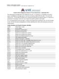
4 Roberto M. Lang, MD, FASE, FESC, received grants from andserves on the speakers bureau and advisory board for Philips Medical Muraru, MD, received research equipment from and served as a consultantfor GE Healthcare. Lawrence Rudski, MD, FASE, holds stock in ASE Members:The ASE has gone green! earn free continuingmedical education credit through an online activity related to this are available for immediate access upon successful completionof the activity. Nonmembers will need to join the ASE to access this greatmember benefit!Drs Lang and Badano co-chaired the Writing requests: American Society of Echocardiography, 2100 Gateway CentreBoulevard, Suite 310, Morrisville, NC 27560 2015 by the American Society of OF CONTENTSI. The Left Ventricle 31. Measurement of LVSize LinearMeasure-ments Volumetric Measure-ments NormalReferenceValues for 2DE NormalReferenceValues for 3DE 6 Recommendation 62.
5 LV Global Systolic Func-tion FractionalShort-ening EF Global LongitudinalStrain (GLS) NormalReferenceValues 7 Recommendations 103. LV Regional Func-tion Segmentation of theLeft Ventricle VisualAssess-ment Regional Wall Motionduring Infarction andIschemia Regional Abnormalitiesin the Absence of Cor-onary Artery Dis-ease QuantificationofRegional Wall MotionUsing Doppler andSTE 11 Recommendations 124. LV Mass 13 Recommendations 16II. The Right Ventricle 165. General Recommenda-tions for RV Quantifica-tion 166. Essential Imaging Win-dows and Views 167. RV Measurements LinearMeasure-ments Volumetric Measure-ments 17 Recommendations 178. RV Systolic Func-tion RIMP TAPSE RV 2D FAC DTI-Derived TricuspidLateral Annular SystolicVelocity RV Strain and Strain Rate 20 Recommendations RV 3D EF 20 Recommendation 20 III.
6 The Left and Right Atria 209. LA Measurements General Considerations for LA Size Linear Dimensions and Area Measurements Volume Measurements Normal Values of LA Measurements 25 Recommendations 2810. Right Atrial measurements 28 Recommendations 28IV. The Aortic Annulus and Aortic Root 2811. The Aortic Annulus 2812. The Aortic Root 3013. Identification of Aortic Root Dilatation 32 Recommendations 32V. The Inferior Vena Cava 32 Notice and Disclaimer 33 References 33 Appendix Measurements Analysis Quantification of Cardiac Chamber size and function is the corner-stone of Cardiac imaging, with echocardiography being the mostcommonly used noninvasive modality because of its unique abilityto provide real-time images of the beating heart, combined with itsavailability and portability.
7 Standardization of the methodologyused to quantify Cardiac chambers is maintained by creating anddisseminating official Recommendations , which when followed bypractitioners provides uniformity and facilitates for echocardiographic Chamber quantificationwere last published in 2005 by the American Society ofEchocardiography (ASE) and the European Association ofEchocardiography (renamed the European Association ofCardiovascular Imaging [EACVI]).1,2 Since then, echocardiographic technology has continuedevolving, with two major developments being real-time three-dimensional (3D) echocardiography (3DE) and myocardial defor-mation imaging. The goal of this document is to provide an updateto the previously published guidelines, as well as recommendationsand reference values, while eliminating the minor discrepancies thatexisted between previous guidelines.
8 The normal values in this up-date include 3DE and myocardial deformation, when , compared with the previous guidelines, this update isbased on considerably larger numbers of normal subjects, compiledfrom multiple databases, to improve the reliability of the most issues covered in this document reflect a broadconsensus among the members of the writing group, one importantissue the group debated was partition values for severity of abnormal-ities. Most often, in addition to describing a parameter as normal orabnormal (reference values), clinical echocardiographers qualify thedegree of abnormality with terms such asmildly,moderately, andAbbreviationsAP= AnteroposteriorASE= American Society ofEchocardiographyBSA= Body surface areaCMR= Cardiac magneticresonanceDTI= Doppler tissue imagingEACVI= EuropeanAssociation of CardiovascularImagingEDV= End-diastolic volumeEF= Ejection fractionESV= End-systolic volumeFAC= Fractional area changeGLS= Global longitudinalstrainI-I= Inner edge to inner edgeIVC= Inferior vena cavaLA= Left atrialL-L= Leading edge to leading edgeLV= Left ventricularMDCT= Multidetectorcomputed tomographyPW= Pulsed-waveRA= Right atrialRIMP= Right ventricular indexof myocardial performanceRV= Right ventricularRWT= Relative wall thicknessSTE= Speckle-trackingechocardiographyTAPSE= Tricuspid annularplane systolic excursionTAVI= Transcatheter aorticvalve implantationTAVR= Transcatheter aorticvalve replacementTEE=
9 Transesophagealechocardiography3D= Three-dimensional3DE= Three-dimensionalechocardiographyTTE= Transthoracicechocardiography2D= Two-dimensional2DE= Two-dimensionalechocardiography2 Lang et alJournal of the American Society of EchocardiographyJanuary 2015severelyabnormal, which reflect the degree to which measurementsdeviate from normal. In addition to providing normative data, itwould be beneficial to standardize cutoffs for severity of abnormalityfor all parameters across echocardiography laboratories, such that thetermmoderately abnormal, for example, would have the same mean-ing universally. However, different approaches may be used for deter-mining cutoff values for the different degrees of abnormality, all ofwhich have significant first approach would be to empirically define cutoffs formild, moderate, and severe abnormalities on the basis of SDs aboveor below the reference limit derived from a group of healthy advantage of this method is that these data readily exist formost echocardiographic parameters.
10 However, this approach isfundamentally flawed. First, not all echocardiographic parametersare normally distributed (or Gaussian), even in a normal , even if a particular parameter is normally distributed innormal subjects, most echocardiographic parameters, whenmeasured in the general population, have a significant asymmetricdistribution in one direction (abnormally large for size or abnormallylow for function parameters). An alternative method would be todefine abnormalities on the basis of percentile values ( , 95th,99th) of measurements derived from a population that includesboth healthy people and those with disease. Although these datawould still not be normally distributed, they would account forthe asymmetric distribution and the range of abnormality presentwithin the general population. The major limitation of this approachis that such population data sets simply do not exist for most echo-cardiographic , an approach that predicts outcomes or prognosis would bepreferred.