Transcription of Rehabilitation Guidelines for Shoulder Arthroscopy
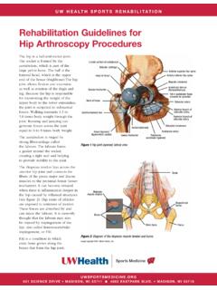
1 621 Science Drive MaDiSon, Wi 53711 Guidelines for Shoulder ArthroscopyThe Shoulder is made up of three bones: the scapula ( Shoulder blade), the humerus (upper arm bone), and the clavicle (collarbone). One part of the scapula, called the glenoid fossa, is coupled with the humerus to make up the socket of the Shoulder (Figure 1). The glenoid is very shallow and flat. The glenoid labrum is a rim of soft tissue that turns the flat surface of the glenoid into a deeper socket that molds to fit the head of the humerus.
2 Another part of the scapula, called the acromium, articulates with the clavicle (collerbone) to make the acromioclavicular (AC) joint . The acromion (Figure 2) itself can be classified as flat (type I), curved (type II), or hooked (type III). The rotator cuff connects the humerus to the scapula. The rotator cuff is formed by the tendons of four muscles: the supraspinatus, infraspinatus, teres minor, and subscapularis (Figure 3). The stability and movement of the Shoulder is controlled primarily by the rotator cuff muscles, with assistance from the ligaments, glenoid labrum and capsule of the Shoulder .
3 Labral tears and rotator cuff tears are often caused by a direct injury to the Shoulder , such as falling on an outstretched hand. However, the labrum and rotator cuff also can become torn from gradual wear and tear of the Shoulder . These tissues can get caught between the glenoid and the humerus or the humerus and the acromion which can cause pain and catching with Shoulder movement. Shoulder Arthroscopy may be performed using instruments (about the size of a pencil which include a camera and other surgical instruments) inserted through small incisions in the Shoulder , to debride massive, irrepairable tears of the labrum and/or rotator ,2 Subacromial impingement occurs when the rotator cuff tendons and/or bursa become trapped between the acromion and the humerus with overhead motion of the This is more likely to occur if the acromion is curved or hooked (Type III)
4 And often leads to pain and limitation of movement at the A subacromial decompression is an arthroscopic procedure performed when an instrument is used to remove some bone on the undersurface of the acromion to create more space for the rotator cuff tendons (Figures 4 and 5). Often there is a bone spur in this region that can pinch against the rotator cuff or bursa (fluid filled sac) causing the pinching or (AC) joint symptoms are another common Shoulder problem, resulting from both direct injury to the AC joint and rotator cuff impingement.
5 A Mumford arthroscopic procedure resects the distal clavicle in cases of Figure 2 Acromion classificationsType IType IIType IIIF igure 1 Shoulder anatomyImage Copyright 2010 UW Health Sports Medicine ViewLong head of bicepShort head of bicepAcromion621 Science Drive MaDiSon, Wi 53711 Guidelines for Shoulder arthroscopyposttraumatic degenerative disease of the AC joint and Shoulder impingement impingement and/or inflammation of the long head of the biceps (Figure 1) can also be a pain generator in the Shoulder .
6 The tendon can often become frayed or partially torn. In some cases the surgeon may release or cut the long head of the bicep near its attachment site to relieve stress and tension, thus eliminating the pain. This is called a biceps tenotomy and can also be done arthroscopically. Rehabilitation is vital to regaining motion, strength and function of the Shoulder after arthroscopic surgery. Initially patients may use a sling for comfort. During this time, range of motion exercises are started to prevent the Shoulder from getting stiff and losing mobility.
7 The Rehabilitation program will gradually progress to more strengthening and control type exercises. General time frames are given for reference to the average, but individual patients will progress at different rates depending on their age, associated injuries, pre-injury health status, Rehabilitation compliance and injury severity. Restrictions or precautions may also be given to protect 4 Pre-operative radiograph of a patient with Shoulder impingement. The arrow indicates the area of the Type III 5 Post-operative radiograph of the same patient in Figure 3.
8 Notice how the Type III acomion (hook) has been shaved off during the subacromial 3 Rotator cuff anatomyImage property of Primal Pictures, Ltd., Use of this image without authorization from Primal Pictures, Ltd. is ViewSupraspinatusInfraspinatusTeres MinorSubscapularisFront View621 Science Drive MaDiSon, Wi 53711 Guidelines for Shoulder arthroscopyPHASE I (usually surgery to 3 weeks after surgery)Appointments Rehabilitation appointments begin 7 days after surgeryRehabilitation Goals Reduce pain and swelling in the post-surgical Shoulder Regain full passive range of motion (PROM) and active assistive range of motion (AAROM)
9 Activation of the stabilizing muscles of the gleno-humeral and scapulo-thoracic jointsPrecautions Avoid activities that may impinge on the denuded bone of the acromion Use sling as needed for comfort Relative rest to reduce inflammationSuggested Therapeutic Exercise Begin 7 days after surgery, sub-maximal Shoulder isometrics for internal rotation/external rotation, flexion/extention and abduction/adduction Shoulder AAROM/PROM: Codman s, pulleys, cane exercises in all planes of motion except horizontal adduction (these should stay relatively pain free) Gentle Shoulder mobilizations as needed Hand gripping Elbow, forearm, and wrist active range of motion (AROM)
10 Cervical spine and scapular AROM Postural exercisesCardiovascular Fitness Walking, stationary bike Avoid running and jumping due to the forces that can occur at landingProgression Criteria The patient can progress to Phase II when they have achieved full PROM and normal (5/5) strength for internal rotation/external rotation with arm at side621 Science Drive MaDiSon, Wi 53711 Guidelines for Shoulder arthroscopyPHASE II (begin after meeting Phase I criteria, usually 4-5 weeks after surgery)Appointments Rehabilitation appointments are once every 1-2 weeksRehabilitation Goals Controlled restoration of AROM Strengthen Shoulder and scapular stabilizers in protected position (0 - 45 abduction)