Transcription of Rehabilitation Protocol: Meniscal Repair
1 Rehabilitation protocol : Meniscal Repair Department of Orthopaedic Surgery Lahey Hospital & Medical Center, Burlington 781-744-8650 Lahey Outpatient Center, Lexington 781-372-7020 Lahey Medical Center, Peabody 978-538-4267 Department of Rehabilitation Services Lahey Hospital & Medical Center, Burlington 781-744-8645 Lahey Hospital & Medical Center, Wall Street, Burlington 781-744-8617 Lahey Danvers 978-739-7400 Lahey Outpatient Center, Lexington 781-372-7060 Meniscal Repair Approved by J. Baumfeld, MD, M. Lemos, MD 12_2013; Compiled by Lauren Scola, DPT, OCS, OMT, Reviewed 2_2015 2 Meniscal Repair : Overview: Meniscal tears occur in different ways. Tears are noted by how they look, as well as where the tear occurs in the meniscus . Common tears include longitudinal, parrot-beak, flap, bucket handle, and mixed/complex.
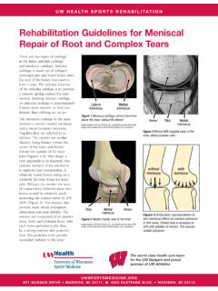
2 The decision by the surgeon to Repair or remove is based primarily on the location of the Meniscal tear. The outer one-third of the meniscus has a rich blood supply. A tear in this red zone may heal on its own, or can often be repaired with surgery. A longitudinal tear is an example of this kind of tear. Tears that exist in the periphery are more likely to heal and as a result often will be repaired, especially in younger patients and in those whose sport or job requires increased stress to the The inner two-thirds of the meniscus lacks blood supply. Without nutrients from blood, tears in the white zone cannot heal. These complex tears are often in thin, worn cartilage. Because the pieces cannot grow back together, tears in this zone are usually surgically trimmed Weight bearing should be limited after Meniscal Repair , and progressed slowly over eight weeks to allow for biological healing.
3 If the Repair is in an area of sufficient vascularity and the fixation is stable, weight bearing is often allowed in fixed extension (brace locked in extension). This position protects the Repair , and allows for controlled axial loading while limiting shear forces that may impede Weight bearing is at the discretion of the surgeon but often is immediate (in full extension) or begins within two weeks progressing to full by the four-week time period. In the first four weeks following the Repair , weight-bearing activities in angles greater than 45 of knee flexion are avoided to allow for healing of the Repair ; while loaded knee flexion beyond 90 is limited for 8 The postoperative Rehabilitation focus on quadriceps strengthening is performed in the open kinetic chain position to avoid loading the meniscus and stressing the fixation or aggravating the In cases of complex Meniscal repairs, weight bearing restrictions can extend to 6-8 weeks due to vascularization and healing.
4 1 Meniscal Tears. In Retrieved 4/3/13, from 2 Brontzman SB, Wilk KE, Clinical Orthopaedic Rehabilitation . Philadelphia, PA: Mosby Inc; 2003:315-319. 3 Current Concepts in Orthopaedic Physical Therapy, 3rd Edition. The Knee: Physical Therapy Patient Management Utilizing Current Evidence Tara Jo Manal, PT, DPT, OCS, SCS; Steve A. Hoffman, PT, ATC, SCS; and Lynne Sturgill, PT, MHS, OCS (Subject Matter Expert: Lisa T. Hoglund, PT, PhD, OCS, Cert MDT) Meniscal Repair Approved by J. Baumfeld, MD, M. Lemos, MD 12_2013; Compiled by Lauren Scola, DPT, OCS, OMT, Reviewed 2_2015 3 Phase I Protective Phase 1 4 Weeks Treatment: Ice, compression, elevation Electrical muscle stimulation WBAT with crutches and brace locked at 0 Motion is limited for the first 7-21 days, depending on the development of scar tissue around the Repair site.
5 Gradual increase in flexion ROM is based on assessment of pain and site of Repair (0 -90 ) Patellar mobilization Scar tissue mobilization Proprioception training with brace locked at 0 Exercises: Quad isometrics, HS isometrics (if posterior horn Repair , no HS exercises until 6 weeks), ankle pumps, Quad sets, heel slides, Hip ABD, Hip ADD Goals Diminish inflammation and swelling Restore ROM (full knee ext, knee flex to 90 , hip and ankle ROM WNL) Reestablish Quad muscle activity-SLR without Quad lag Full scar mobility Full patellar mobility Precautions WB status: WBAT with crutches and brace locked at 0 ROM Restrictions: Passive ROM 0 -90 Recommended Restrictions: avoid pivoting and varus/valgus stress, no flexion beyond 90 Meniscal Repair Approved by J.
6 Baumfeld, MD, M. Lemos, MD 12_2013; Compiled by Lauren Scola, DPT, OCS, OMT, Reviewed 2_2015 4 Phase II Intermediate Phase Weeks 4 6 Treatment: Immobilizer D/C per surgeon Progress PRE s for hip, knee, ankle Progress WB flex 45 -90 Continue proprioceptive training Exercises: heel raises, mini-squats (less than 90 knee flexion), stationary bike (no resistance) Goals Diminish inflammation and swelling Restore Full AROM Reestablish Quad muscle activity Criteria to progress to next phase: ROM 0 -90 , no change in pain or effusion, Quad control (MMT 4/5) Precautions WB status: WBAT with crutches and brace locked at 0 ROM Restrictions: Passive ROM 0 -90 Recommended Restrictions: Avoid pivoting and varus/valgus stress, no flex beyond 90 Meniscal Repair Approved by J.
7 Baumfeld, MD, M. Lemos, MD 12_2013; Compiled by Lauren Scola, DPT, OCS, OMT, Reviewed 2_2015 5 Phase III Week 6-12 Treatment: Begin loaded flex beyond 90 at 8 weeks Exercises: Progress PRE s, stationary bike, Quad set, heel slides, SLR, SAQ, heel raises, side lying hip ABD, standing HS curl, wall slides with knee flex <90 , squat to chair, seated leg press, step up/down, flexibility exercise (HS, Quad, Gastroc stretching) Phase IV Weeks 12-16 Treatment: Continue all exercises Increase plyometrics and pool program (if available) Initiate running program Sport specific drills Emphasize plyometrics, jumping, cutting Goals Restore full AROM Regain full muscle strength Prepare patients for advances exercises Criteria to progress to next phase.
8 Full, pain free ROM, no pain or tenderness, SLR without lag, gait without device, brace unlocked Precautions WB status: FWB D/C crutches and brace ROM restrictions: WB flex 0 -90 Recommended Restrictions: Avoid patellofemoral overload, avoid squatting, avoid pivoting or twisting on knee Goals Regain full muscle strength Increase power and endurance Prepare for return to unrestricted activities Work on cardiovascular conditioning Sport-specific training Precautions WB status: FWB ROM Restrictions: No restrictions Recommended Restrictions: Avoid patellofemoral overload, avoid squatting, avoid pivoting or twisting on knee Meniscal Repair Approved by J. Baumfeld, MD, M. Lemos, MD 12_2013; Compiled by Lauren Scola, DPT, OCS, OMT, Reviewed 2_2015 6 Exercises: Progress PRE s, flexibility exercises, mini-squats, step up/down, lateral step-ups, stationary bike, swimming (no from kick), pool running, balance, backward walking, plyometrics Phase V Weeks 16 onward Treatment: Return to Running Progression: Light running on soft, level surface per MD Need full ROM, good strength and no swelling to run safely Start with running 10 minutes, 3 times per week for first 2 pain free with running, can increase running time by 1 minute per session for max 30 minutes Speed and Agility Running Program for Return to Sport.
9 Straight ahead running phase Direction change running phase Unrestricted direction change Goals Safely recondition the injured area for the demands of sports activity Criteria for discharge from skilled therapy: Non-antalgic gait pattern Pain free full ROM No palpable edema LE strength at least 4/5 Independent HEP Age appropriate balance and proprioception abilities Precautions WB status: FWB ROM Restrictions: No restrictions Recommended Restrictions: Avoid patellofemoral overload, avoid squatting, avoid pivoting or twisting on knee Meniscal Repair Approved by J. Baumfeld, MD, M. Lemos, MD 12_2013; Compiled by Lauren Scola, DPT, OCS, OMT, Reviewed 2_2015 7 Rehabilitation protocol for Meniscal Repair Rehabilitation Guidelines: Summary Table Post op Phase/Goals Therapeutic Exercise Precautions Phase I Weeks 1-4 Goals: Diminish inflammation and swelling Restore ROM (full knee ext, knee flex to 90 , hip and ankle ROM WNL) Reestablish Quad muscle activity-SLR without Quad lag Full scar mobility Full patellar mobility Ice, compression, elevation Electrical muscle stimulation WBAT with crutches and brace locked at 0 Motion is limited for the first 7-21 days, depending on the development of scar tissue around the Repair site.
10 Gradual increase in flexion ROM is based on assessment of pain and site of Repair (0 -90 ) Patellar mobilization Scar tissue mobilization Proprioception training with brace locked at 0 Exercises: Quad isometrics, HS isometrics (if posterior horn Repair , no HS exercises until 6 weeks), ankle pumps, Quad sets, heel slides, Hip ABD, Hip ADD WB status: WBAT with crutches and brace locked at 0 ROM Restrictions: Passive ROM 0 -90 Recommended Restrictions: avoid pivoting and varus/valgus stress, no flex beyond 90 Phase II Weeks 4-6 Goals: Diminish inflammation and swelling Restore full AROM Reestablish Quad muscle activity Criteria to progress to next phase: ROM 0 -90 no change in pain or effusion, Quad control (MMT 4/5) Immobilizer D/C per surgeon Progress PRE s for hip, knee, ankle Progress WB flex 45 -90 Continue proprioceptive training Exercises: heel raises, mini-squats (less than 90 knee flex), stationary bike (no resistance) WB status: WBAT with crutches and brace locked at 0 ROM Restrictions: Passive ROM 0 -90 Recommended Restrictions: avoid pivoting and varus/valgus stress, no flex beyond 90 Phase III Weeks 6 - 12 Goals Restore full AROM Regain full muscle strength Prepare patients for advances exercises Criteria to progress to next phase: full, pain free ROM, no pain or tenderness, SLR without lag, gait without device, brace unlocked Treatment: Begin loaded flex beyond 90 at 8 weeks Exercises.