Transcription of Reproduction in camels - A review
1 -28- CHAPTER 3. PROBLEMS OF Reproduction . B. Musa and H. Merkt The problems of Reproduction in the camel are not extensively investigated as for example in the bovine. The information collected on these problems is derived mainly from questioning the camel owners, slaughterhouse material and very limited clinical and farm observations. Early embryonic mortality The incidence of early embryonic death seems to be high in the camel. It was found that twinning occurs in about percent of 491 single births reported by Musa & Abu Sineina (1976 a). In the same study two and three corpora lutea were found in percent and percent respectively. The reason for these high prenatal losses is still open for more investigation (see ). Yagil (1985) claims that one of the causes of foetal death is the strong inbreeding in the herds. Reproductive diseases Abortion and stillbirth These are known to occur in camels .
2 The incidence of brucellosis in camels in different countries is given in Table 3. It seems to related to breeding and husbandry practices. In Africa Brucella melitensis was found to be the causative agent while in the USSR the infection was found to be due to Br. abortus. The organisms were isolated in the different countries with variable incidence of success. It is claimed that young camels are resistant up to the age of 11. months and that they contract the disease from the dams on subsequent calving. If this is the case, then separation of young camels at 7 to 8. months of age from positive dams might help in the control programme (Higgins, 1986). The role played by brucellosis in the overall picture of abortion in camels is not quantified. It appears that other important endemic diseases play a significant role in the overall incidence of abortion. Trypanososmiasis leading to general debility and abortion is an important disease.
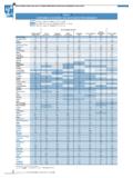
3 Pasteurellosis and salmonellosis are also considered as causes of abortion in camels . Other causes of abortion include febrile conditions such as pneumonia and camel pox, or nervous excitement (Mukasa-Mugerwa, 1981). - 29 - Table 3: Incidence of brucellosis in camel populations in different countries (after Higgins, 1986). Country Infection Rate percent USSR Chad Ethiopia Egypt - Sudan - North-Eastern Kenya Nigeria India Tunisia - - 30- Diseases of the female reproductive tract Examination of abattoir material provided some information about some of the diseases that could be encountered in the reproductive tract. Although this could be considered as biased information when referring to populations of camels in general, it could however provide useful information about the existence of these diseases. These diseases include: pyometra, bursal and ovarian adhesions, endometritis associated with a partially involuted uterus, and cystic ovarian degeneration (Mukasa-Mugerwa, 1981).
4 Dystocia The incidence of camel dystocia appears to be very low (Arthur et al., 1985 a). The foetal component of dystocia includes: carpal flexion, lateral deviation of the head and hock and hip flexion. Posterior presentation is uncommon. Foetopelvic disproportion, monstrosities and transverse presentations are rare. On the maternal side, uterine inertia occurs to a small extent. In dealing with dystocia in the camel it was found that head and limb extension is more difficult to achieve than in the cow. However, the camel foetus survives dystocia better than the equine foetus and the camel is a good subject for caesarean section. Also foetotomy using Thygensen's embryotome is possible when necessary. Ceasarean section could be performed on the left flank using xylazine sedation and local regional or infiltration anaesthesia. A camel, 17 hours in dystocia, delivered a live foetus by caesarean section (Arthur et al.)
5 , 1985 a). Vaginal prolapse This is seen clinically in pregnant camels that were well fed with limited exercise. In most of the cases it does not interfere with the pregnancy in question. The condition could be treated successfully by the B hrer technique (Arthur et al., 1985 a). Other problems of Reproduction The fertility is defined as the ability of the male and female to produce viable germ cells, mate and conceive and subsequently give birth to living young (Mukasa-Mugerwa, 1981). A significant aid to establish precise figures for fertility is record keeping and good management. Unfortunately both are missing under traditional camel raising systems. Existing information indicates that fertility is unlikely to be higher than 50 percent in pastoral herds and that under improved ranch conditions it could be up to 65 percent (Mukasa-Mugerwa, 1981). - 31 - In Saudi Arabia Arthur et al. (1985 b) reported a 80 - 90 percent fertility rate and that sterility is about 1 percent.
6 Yagil (1985). observed up to 100 percent fertility. Poor nutrition and poor grazing are a cause of reduced sexual activity in both females and males. Debilitating diseases such as trypanosomiasis, tuberculosis, mange, pleuropneumonia and heavy parasitism all compromise the fertility rate in the camel. Endocrine factors including insufficient gonadotropins to enhance follicular development and subsequent ovulation may also contribute to fertility rate in camels . Concerning the bacterial flora of the female genitalia in the camel, Zaki & Mousa (1965) isolated corynebact., anthrocoids, micrococci, sarcina, gaffkya and gram negative becilli from the normal genital tract of pregnant and non-pregnant slaughtered animals. Almost the same spectrum of germs was found by Eidarous et al., (1983), however including E. coli and Staph. epidermidis. Nawito (1973) recorded the bacteriological findings in the uteri of 2075 one-humped camels of unknown history from the Cairo abattoir.
7 In 94. cases ( percent) clinical symptoms such as asbcesses in the uterus, catarrhal endometritis, haemorrhagic endometritis, pyometra and pyometra with macerated foeti were found. Micrococcus pyogenes var. aureus played the predominant role. Furthermore, Micrococcus pyogenes var. albus, beta-haemolytic streptococci, E. coil and Pseudomonas aeruginosa could be isolated from the uteri of animals with clinical symptoms. Reproductive problems in the male There is very little information on reproductive problems in the male camel. Phimosis, paraphimosis, orchitis and testicular hypoplasia were clinically observed. Abdel-Raouf (1965) described a case of bilateral testicular hypoplasia in a dromedary. The seminiferous tubules were divided into four types according to the degree of development. Microscopic examination revealed that the smaller left testicle contained a larger number of underdeveloped tubules than the right one.
8 - 32 - Burgemeister (1974) observed a high incidence of incest breeding in camel herds in Tunisia. It is well known that inbreeding can cause alterations in the male genital tract such as hypoplasia. We found cases of unilateral cryptorchidism both in a live animal and in material from abattoirs (Fig. 3, 4). Furthermore, a case of subfertility was found due to pronounced asthenozoospermia in the semen of a male camel with normally developed and clinically healthy testicles. Shawki et al. (1983) reported the incidence of filariosis among Egyptian camels to be 5 percent. The histological changes of both the testis and epididymis affected with filariosis reveal fibrosis of the tunica albuginea. The consistency of the compromised testis was soft with areas of hardness. There were degenerative changes and necrosis of the seminiferous tubules and epididymal ductus. The arterioles were thickened, dilated and engorged with microfilaria.
9 These pathological changes were due to occlusion of the arteries and arterioles with larvae, thus reducing circulation to the testis and the epididymis. Such conditions may induce sterility in camels . With regard to the age, the minimal incidence of testicular degeneration ( percent) was found between the age of 4 - 6 years, while the maximum values (50 percent) were present in senile camels over 20 years of age. Marked decline in the blood levels of thyroxine, carotene and vitamin A were found in camels with moderate and advanced testicular degenerations. Hemeida et al. (1985 b) mentioned that there were different degrees of testicular degenerations. These changes exert a profound influence on the sperm production rate. Other testicular abnormalities mentioned by Hemeida et al. (1985 c) were: testicular hypoplasia ( percent), cryptorchidism ( percent), orchitis ( percent), necrosis ( percent), hydrocele ( percent) and seminoma ( percent).
10 Filarial orchitis and funiculitis due to dipetalonema evansi was the most common abnormality ( percent). The bacterial flora of the male and female genital system of the camel has been examined by Eidarous et al. (1983). They found that Staph. epidermidis, anthracoids, E. coli, micrococci and Gaffkya were the most prevalent bacteria in the male genital tract. The prepuce and urethra were the organs most inhabited by microf lora. Staph. aureus, B. proteus, Pseudomonas aeruginosa, C. bovis and streptococci were isolated only from the prepuce. The percentage of male genitalia in which no microbes were found was about 42 percent. - 33 - CHAPTER 4. RESEARCH NEEDS FOR IMPROVING CAMEL PRODUCTIVITY. B. Musa and H. Merkt Improvement of low fertility rates The low fertility rates in camels constitute an obstacle in camel Reproduction and hence in camel production. In order to increase offtake rate in any population of camels , one has to improve the fertility rate in that population.