Transcription of Review paper Biomechanics of the cervical spine. I: Normal ...
1 Clinical Biomechanics 15 (2000) 633 648. Review paper Biomechanics of the cervical spine . I: Normal kinematics Nikolai Bogduk a,*, Susan Mercer b a Newcastle Bone and Joint Institute, University of Newcastle, Royal Newcastle Hospital, Level 4, David Maddison Building, Newcastle, NSW 2300, Australia b Department of Anatomy, University of Otago, Dunedin, New Zealand Abstract This Review constitutes the rst of four reviews that systematically address contemporary knowledge about the mechanical behavior of the cervical vertebrae and the soft-tissues of the cervical spine , under Normal conditions and under conditions that result in minor or major injuries. This rst Review considers the Normal kinematics of the cervical spine , which predicates the appreciation of the Biomechanics of cervical spine injury.
2 It summarizes the cardinal anatomical features of the cervical spine that determine how the cervical vertebrae and their joints behave. The results are collated of multiple studies that have measured the range of motion of individual joints of the cervical spine . However, modern studies are highlighted that reveal that, even under Normal conditions, range of motion is not consistent either in time or according to the direction of motion. As well, detailed studies are summarized that reveal the order of movement of individual vertebrae as the cervical spine exes or extends. The Review concludes with an account of the location of instantaneous centres of rotation and their biological basis. Relevance The facts and precepts covered in this Review underlie many observations that are critical to comprehending how the cervical spine behaves under adverse conditions, and how it might be injured.
3 Forthcoming reviews draw on this information to explain how injuries might occur in situations where hitherto it was believed that no injury was possible, or that no evidence of injury could be detected. 2000 Elsevier Science Ltd. All rights reserved. Keywords: cervical spine ; Biomechanics ; Movements; Anatomy 1. Introduction therefore, predicated by the anatomy of the bones that make up the neck and the joints that they form. Amongst its several functions, the head can be re- garded as a platform that houses the sensory apparatus for hearing, vision, smell, taste and related lingual and 2. Functional anatomy labial sensations. In order to function optimally, these sensory organs must be able to scan the environment For descriptive purposes, the cervical spine can be and be delivered towards objects of interest.
4 It is the divided and perceived as consisting of four units, each cervical spine that subserves these facilities. The cervical with a unique morphology that determines its kine- spine constitutes a device that supports the sensory matics and its contribution to the functions of the platform, and moves and orientates it in three-dimen- complete cervical spine . In anatomical terms the units sional space. are the atlas, the axis, the C2 3 junction and the re- The movements of the head are executed by muscles maining, typical cervical vertebrae. In metaphorical, but the type of movements possible depend on the shape functional terms these can be perceived as the cradle, the and structure of the cervical vertebrae and interplay axis, the root, and the column.
5 Between them. The kinematics of the cervical spine are, The cradle The atlas vertebra serves to cradle the occiput. Into *. Corresponding author. its superior articular sockets it receives the condyles of E-mail address: (N. Bogduk). the occiput. The union between the head and atlas, 0268-0033/00/$ - see front matter 2000 Elsevier Science Ltd. All rights reserved. PII: S 0 2 6 8 - 0 0 3 3 ( 0 0 ) 0 0 0 3 4 - 6. 634 N. Bogduk, S. Mercer / Clinical Biomechanics 15 (2000) 633 648. through the atlanto-occipital joints, is strong, and allows only for nodding movements between the two struc- tures. In all other respects the head and atlas move and function essentially as one unit. The stability of the atlanto-occipital joint stems largely from the depth of the atlantial sockets.
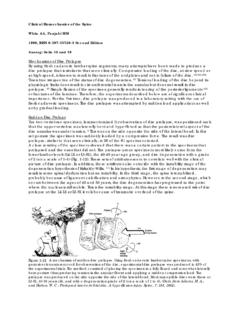
6 The side walls of the sockets prevent the occiput from sliding Fig. 2. Right lateral views of axial rotation of the atlanto-occipital sideways; the front and back walls prevent anterior and joints. Rotation requires forward translation of one condyle and posterior gliding of the head, respectively. The only backward translation of the other. Translation is possible only if the physiological movements possible at this joint are ex- condyles rise up the respective walls of the atlantial sockets. As a re- sult, the occiput rises relative to its resting position (centre gure). ion and extension, nodding. These are possible be- cause the atlantial sockets are concave whereas the occipital condyles are convex. against the posterior wall of its socket.
7 For the head to Flexion is achieved by the condyles rolling forwards rotate, the condyles must rise up their respective walls. and sliding backwards across the anterior walls of their Consequently, the occiput must separate from the atlas sockets (Fig. 1). If the condyles only rolled, they would (Fig. 2). This separation is resisted by tension in the roll up and over the anterior wall of their sockets. Axial capsules of the atlanto-occipital joints. As a result, the forces exerted by the mass of the head or the muscles range of motion possible is severely limited. Lateral causing exion prevent this upward displacement and exion is limited by similar mechanisms. For lateral cause the condyles to slide downwards and backwards exion to occur the contralateral condyle must lift out of across the concave surface of the socket.
8 Thereby the its socket, which engages tension in the joint capsule. condyles remain within their sockets, and the composite movement is a rotation, or a spin, of each condyle across The axis the surface of its socket. A converse combination of movements occurs in extension. This combination of Carrying the head the atlas sits on the atlas, with the roll and contrary glide is typical of condylar joints. weight being borne through the lateral atlanto-axial The ultimate restraint to exion and extension of the joints. After weight-bearing, the cardinal function of the atlanto-occipital joint is impaction of the rim of the atlanto-axial junction is to permit a large range of axial socket against the base of the skull.
9 Under Normal rotation. This movement requires the anterior arch of conditions, however, exion is limited by tension in the the atlas to pivot on the odontoid process and slide posterior neck muscles and by impaction of the sub- around its ipsilateral aspect; this movement being mandibular tissues against the throat. Extension is lim- accommodated at the median atlanto-axial joint ited by the occiput compressing the suboccipital (Fig. 3(A)). Meanwhile, at the lateral atlanto-axial joint muscles. the ipsilateral lateral mass of the atlas must slide back- Axial rotation and lateral exion are not physiologi- wards and medially while the contralateral lateral mass cal movements of the atlanto-occipital joints. They must slide forwards and medially (Fig.)
10 3). cannot be produced in isolation by the action of mus- Radiographs of the lateral atlanto-axial joints belie cles. But they can be produced arti cially by forcing the their structure. In radiographs the facets of the joint head into these directions while xing the atlas. Axial appear at, suggesting that during axial rotation the rotation is prohibited by impaction of the contralateral lateral atlanto-axial joints glide across at surface. But condyle against the anterior wall of its socket and si- radiographs do not reveal cartilage. The articular car- multaneously by impaction of the ipsilateral condyle tilages both of the atlantial and the axial facets of the Fig. 1. Right lateral views of exion and extension of the atlanto-oc- cipital joints.