Transcription of Review SLAP lesions: Anatomy, clinical presentation, MR ...
1 European Journal of Radiology 68 (2008) 72 87 ReviewSLAP lesions: anatomy , clinical presentation , MR imaging diagnosisand characterizationDebra Changa,b,c, , Aurea Mohana-Borgesa,b, Maya Borsoa,b, Christine B. Chunga,baUniversity of California San Diego, Department of Radiology, 200 W. Arbor Drive, San Diego,CA 92103, United StatesbVA Healthcare System San Diego, Department of Radiology, 3350 La Jolla Village Drive,La Jolla, CA 92161, United StatescMedRay Imaging and Fraser Health Authority, Vancouver, BC, CanadaReceived 8 February 2008; received in revised form 9 February 2008; accepted 19 February 2008 ABSTRACTS uperior labral anterior posterior ( slap ) tears are an abnormality of the superior labrum usually centered on the attachment of the long headof the biceps tendon.
2 Tears are commonly caused by repetitive overhead motion or fall on an outstretched arm. slap lesions can lead to shoulderpain and instability. clinical diagnosis is difficult thus imaging plays a key diagnostic role. The normal anatomic variability of the capsulolabralcomplex can make slap lesions a diagnostic challenge. Concurrent shoulder injuries are often present including rotator cuff tears, cystic changesor marrow edema in the humeral head, capsular laxity, Hill-Sachs or Bankart lesion . The relevant anatomy , capsulolabral anatomic variants, primaryand secondary findings of slap tears including MR arthrography findings, types of slap lesions and a practical approach to labral lesions arereviewed. 2008 Elsevier Ireland Ltd. All rights :Superior labral anterior posterior lesion slap lesion ; Glenoid labrum; Shoulder anatomy ; MR arthrography; presentation , etiology, and physical of the capsulolabral variants of the superior and anterosuperior normal labral variants from of slap practical approach to slap labral anterior posterior ( slap ) tears are a com-mon cause of labral pathology, leading to shoulder pain and Corresponding author at: University of California San Diego, Departmentof Radiology, 200 W.
3 Arbor Drive, San Diego, CA 92103, United : +1 619 471 0514; fax: +1 619 471 Chang).instability. A slap lesion is an acquired abnormality of thelabrum, usually centered on the attachment of the long head ofbiceps tendon. It can extend to involve the anterior and poste-rior labrum, as well as the surrounding anatomic structures. Thesuperior labrum is functionally important as an anchor for theinsertion of the biceps tendon on the glenoid rim. Andrews et al.[1]first described detachment and fraying of the anterior supe-rior labrum, which may be accompanied by partial tearing of the0720-048X/$ see front matter 2008 Elsevier Ireland Ltd. All rights Chang et al. / European Journal of Radiology 68 (2008) 72 8773biceps tendon, in a group of high-level throwing athletes.
4 Theterm slap was subsequently coined by Snyder et al.[2], whodescribed a similar but more global injury of the superior labrumthat extended anterior and posterior to the biceps tendon. Diag-nosis of a slap lesion can be difficult clinically, and imagingplays a key role in detecting a slap studies have reported a prevalence of slap lesions of Snyder et al.[3]analyzed 700 shoul-der arthroscopies and found a slap incidence of Maffetet al.[4]identified 84 slap -type lesions in 712 consecutiveshoulders arthroscopies, constituting an incidence rate of mechanisms of injury include repetitive overhead armmotion, found in throwers and swimmers, as well as a fall onthe outstretched arm in the acute Review will familiarize the reader with the anatomy andanatomic variations of the capsulolabral complex of the gleno-humeral joint, as we Review the clinical presentation and etiologyof slap pathology, and describe the MR imaging techniquesand findings that aid in the diagnosis and characterization ofSLAP clinical presentation , etiology, and physical examThe clinical diagnosis of a slap lesion is difficult.
5 Non-specific shoulder pain, particularly with overhead or cross-bodymotion, is the most common clinical presentation . Additionalsymptoms include popping, clicking, catching, weakness, stiff-ness and instability. The majority of patients present withconcurrent shoulder injuries. In a retrospective Review of 140arthroscopically-proven slap lesions by Snyder et al.[3], thereported incidence of associated intra-articular disease included29% with partial rotator cuff tears, 11% with full rotator cufftears, 22% with Bankart lesions and 10% with history may involve a traction injury, direct traumato the shoulder, or fall on an outstretched arm. Frequently, noantecedent injury or activity is reported. On physical exam, thepatient may have increased shoulder laxity and positive findingswith many provocative shoulder tests.
6 No single physical testor sign is specific for slap lesions and physical findings canbe confusing due to associated lesions ( rotator cuff tears).The clinical diagnosis of a slap lesion is difficult and imagingplays a key role in , a cadaveric study has confirmed the peel-back the-ory of slap lesions. In the abducted and externally rotatedshoulder, the biceps tendon assumes a more vertical and posteri-orly directed orientation, which transmits a force to the superiorlabrum, causing it to peel off the glenoid[5]. Common mech-anisms of injury include microtrauma secondary to repetitiveoverhead arm motion, and direct trauma due to falling on anoutstretched hand. Repetitive overhead arm motions, such asthose used in throwing and swimming, are thought to causeinjury secondary to traction on the arm due to sudden pulling,throwing or other overhead motion[4].
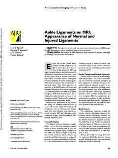
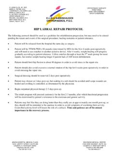
7 Additional findings inrepetitive overhead motion injury include, undersurface rotatorcuff tears, cystic change in the humeral head related to pos-terosuperior impingement and capsular laxity. Falling on anFig. 1. Labral localization. (A) Clockface superimposed upon the glenoid inthe sagittal plane allows precise localization of labral pathology. Note that the 3o clock position is always anterior, 9 o clock posterior, 12 o clock superior and6 o clock inferior, regardless of right versus left side. (B) Labral localizationcan also be performed in a slightly more general fashion by dividing the glenoidinto Chang et al. / European Journal of Radiology 68 (2008) 72 87 Fig. 2. Transitional zone. Axial T1-weighted fat-suppressed MR arthrogramimage demonstrates intermediate signal undercutting the contour of the posteriorsuperior glenoid labrum (arrowhead) representing the histologic transitional areafrom labral fibrocartilage to hyaline articular cartilage of the arm causes injury secondary to a compressive forceapplied to the shoulder, usually with the shoulder abducted andslightly anteriorly flexed[2].
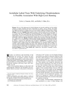
8 This mechanism can result in mar-row edema secondary to impaction of the humeral head againstthe glenoid. If an associated anterior dislocation is present, aHill-Sachs deformity and a Bankart lesion may anatomy of the capsulolabral complexThe labrum is a fibrocartilaginous structure that forms acuff around the periphery of the glenoid. The labrum deepensthe glenoid fossa and provides shoulder stability. It serves asthe attachment for the glenohumeral ligaments and the tendonof the long head of the biceps muscle to the glenoid. Anteri-Fig. 3. Buford complex. Axial proton density-weighted fat-suppressed MRimage demonstrates absence of the anterior superior labrum (straight arrow)with a thickened cord-like MGHL (curved arrow).orly, the labrum blends with the anterior band of the inferiorglenohumeral ligament (IGHL).
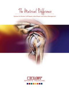
9 Superiorly, it blends with thebiceps tendon and superior glenohumeral ligament (SGHL). Thelabrum is variable in shape, size and attachment to the glenoid[6]. The labrum is usually rounded or triangular. Its superiorportion is more loosely attached and more mobile than the restof the labrum. This normal laxity can be confused with slap lesions. The labrum normally displays low signal on all descriptive purposes, labral position is localized by super-imposing the face of a clock onto the surface of the glenoid, andby convention 3 o clock is anterior, 9 o clock is posterior, 12o clock is superior, and 6 o clock is inferior. An alternative isFig. 4. Sublabral recess or sulcus. Coronal (A) and axial (B) T1-weighted fat-suppressed MR arthrogram images demonstrate a linear, well-defined spacebetween the superior labrum and the adjacent osseous glenoid that parallels theglenoid margin.
10 The sulcus is usually confined to the superior labrum at the 11 1o clock position at the site of attachment of the long head of biceps Chang et al. / European Journal of Radiology 68 (2008) 72 8775to divide the labrum into six segments superior, anterosupe-rior, anteroinferior, inferior, posteroinferior, and posterosuperior(Fig. 1A and B).The three-glenohumeral ligaments are focal thickenings ofthe anterior joint capsule and can have significant variation inanatomy. The superior glenohumeral ligament can arise fromthe anterosuperior labrum, the biceps tendon attachment, or themiddle glenohumeral ligament (MGHL). The SGHL is orientedin an axial plane extending from the superior glenoid to thelesser tuberosity, and it blends with the coracohumeral SGHL is a minor contributor to glenohumeral stability.