Transcription of RHABDOMYOLYSIS: PREVENTION AND TREATMENT
1 DISCLAIMER: These guidelines were prepared jointly by the surgical critical care and Medical critical care Services at Orlando Regional Medical Center. They are intended to serve as a general statement regarding appropriate patient care practices based upon the available medical literature and clinical expertise at the time of development. They should not be considered to be accepted protocol or policy, nor are intended to replace clinical judgment or dictate care of individual patients. EVIDENCE DEFINITIONS Class I: Prospective randomized controlled trial. Class II: Prospective clinical study or retrospective analysis of reliable data. Includes observational, cohort, prevalence, or case control studies. Class III: Retrospective study.
2 Includes database or registry reviews, large series of case reports, expert opinion. Technology assessment: A technology study which does not lend itself to classification in the above-mentioned format. Devices are evaluated in terms of their accuracy, reliability, therapeutic potential, or cost effectiveness. LEVEL OF RECOMMENDATION DEFINITIONS Level 1: Convincingly justifiable based on available scientific information alone. Usually based on Class I data or strong Class II evidence if randomized testing is inappropriate. Conversely, low quality or contradictory Class I data may be insufficient to support a Level I recommendation. Level 2: Reasonably justifiable based on available scientific evidence and strongly supported by expert opinion.
3 Usually supported by Class II data or a preponderance of Class III evidence. Level 3: Supported by available data, but scientific evidence is lacking. Generally supported by Class III data. Useful for educational purposes and in guiding future clinical research. 1 Approved 02/01/2005 Revised 10/07/2009, 07/27/2015, 07/24/2018 2018 RHABDOMYOLYSIS: PREVENTION AND TREATMENT SUMMARY Rhabdomyolysis (RM) was originally described in patients with crush injury, but non-traumatic causes are also common. A high index of suspicion is necessary to allow prompt recognition and TREATMENT to avoid the development of acute renal failure (ARF) and need for hemodialysis. Classically, RM is treated with fluid administration and diuretics as well as bicarbonate therapy in an attempt to alkalinize the urine.
4 More recently, these adjuncts have come into question and it appears that prompt recognition and appropriate volume replacement is all that is needed to avoid renal deterioration. INTRODUCTION RM is the dissolution muscle and release of potentially toxic intracellular components into the systemic circulation (1). RM has the potential to cause myoglobinuric ARF in 10-15% of such patients. Overall, 10-15% of ARF in the United States is from RM. Creatine phosphate (CP) is found in striated muscle and is a reservoir of high-energy phosphate bonds. Creatine phosphokinase (CPK) catalyzes the regeneration of adenosine triphosphate (ATP) from the combination of CP with adenosine diphosphate (ADP). In RM, muscle cells die and release the CPK enzyme into the bloodstream.
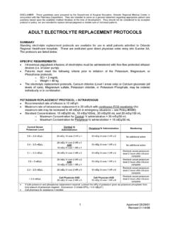
5 Myoglobin (MG) is an oxygen binding protein that composes 1-3% of the dry weight of skeletal muscle. It has a high affinity for oxygen accepting oxygen molecules from hemoglobin in the bloodstream. With RECOMMENDATIONS Level 1 None Level 2 Lactated Ringers solution is the fluid of choice when attempting to maintain adequate urinary output ( mL/kg) in patients with rhabdomyolysis (RM). Level 3 In patients with RM with low urine output unresponsive to fluid administration alone or a creatinine kinase of 10,000 u/L, alkalinization of the urine and addition of mannitol is warranted. In patients with RM, it is important to minimize other potential renal insults (such as nephrotoxic antibiotics, intravenous contrast media, ACE inhibitors, NSAIDS, ).
6 Serial CK measurements to monitor the resolution of RM are not warranted after the zenith is reached; 2 Approved 02/01/2005 Revised 10/07/2009, 07/27/2015, 07/24/2018 2018 muscle damage, free MG in the blood leads to myoglobinemia. Normally, low levels are well tolerated and are cleared by the reticuloendothelial system. At high levels, however, binding and normal clearing mechanisms become saturated, eventually leading to myoglobinuria and the potential for renal injury and ARF. Myoglobinuria is the presence of MG in the urine. The urine is found to be positive for blood despite the absence of erythrocytes on microscopic examination. MG contains iron, the toxic effects of which are described below.
7 MG also has the potential to release vasoactive agents such as platelet activating factor and endothelins which may lead to renal arteriolar vasoconstriction, thus worsening renal function. MG appears first in the plasma, but is rapidly cleared within 24 hours. CPK appears a few hours later than MG, reaches its peak value within the first 24 hours, and remains at such levels for several days. CPK is considered to be a more useful marker for the diagnosis and assessment of the severity of muscular injury due to its delayed clearance from the plasma. A prerequisite for the development of this disease process is muscle injury, the causes of which are numerous and outlined below. While low levels of ischemia (< hours) are typically well tolerated, as the ischemic time lengthens irreversible muscle damage occurs allowing the release of toxic metabolic byproducts.
8 Reperfusion after a period of ischemia contributes to localized tissue edema mediated by leukocytes, leukotrienes and inflammatory mediators. Cell membranes are damaged, cellular contents leak, and intracellular ATP, the main fuel for cellular membrane pumps, is depleted worsening cellular homeostasis. Another problem is the development of intracellular hypercalcemia leading to the activation of intracellular autolytic enzymes that damage cell membranes leading to the cells vulnerability to oxygen free radicals with reperfusion. There are various causes of RM: vascular interruption, ischemia-reperfusion, crush injury, improper patient positioning, alcohol ingestion, seizures, extreme exercise, electrical injury, infection, hyperthermia, and steroids and neuromuscular blockade (especially in combination).
9 With heightened suspicion for this disorder, non-traumatic causes are being seen with increasing frequency. A special group that has recently been seen to be at risk is bariatric surgery patients. RM is increasingly being seen in clinical practice as the popularity of bariatric surgery is gaining momentum. Several longitudinal studies have found rates of RM after bariatric surgery ranging from 7-77%. A rare syndrome leading to RM specifically related to these patients is known as gluteal compartment syndrome. PHYSIOLOGICAL BASIS OF TREATMENT MODALITIES The most important component with regard to the TREATMENT of patients with RM is the ability to recognize the disease process in a timely fashion to prevent the consequences of myoglobinuria.
10 Worsening renal function as evidenced by increasing blood urea nitrogen (BUN) and creatinine, oliguria, classic tea colored urine , and an elevated serum CPK level all but make the diagnosis. Other findings include hypocalcemia, hyperkalemia and the potential for cardiac toxicity, hyperuricemia, hyperphosphatemia, lactic acidosis, and disseminated intravascular coagulation (DIC) from thromboplastin release. The cornerstone of TREATMENT is aggressive volume resuscitation and expansion of the extracellular fluid compartment. Other modalities described include the use of bicarbonate in an attempt to alkalinize the urine, mannitol, and iron chelators (deferoxamine). Prompt and aggressive restoration of volume is essential and critical to prevent progression to ARF and the need for renal replacement therapy and its inherent cost, morbidity, and mortality.