Transcription of SELECTIVE AND DIFFERENTIAL MEDIA - Youngstown State …
1 Microbiology Laboratory (BIOL 3702L) Page 1 of 7. SELECTIVE AND DIFFERENTIAL MEDIA . Principle and Purpose Microorganisms are generally studied after being grown on or in a microbiological medium. There is an expansive variety of useful MEDIA for this purpose including many types of general growth MEDIA that support a wide spectrum of organims. Examples include defined MEDIA , such as glucose-salts, as well as rich, chemically complex MEDIA such as brain heart infusion agar and tryptic soy broth. However, these general growth MEDIA may permit the growth unwanted microorganisms from clinical or environmental samples that are not relevant to the purpose of the microbiological examination.
2 To help resolve this issue, the components of differnt MEDIA are sometimes adjusted to enhance the growth of particular organisms or, alternatively, to retard the growth of unwanted microbes. In the clinical laboratory, this ability to preferentially grow one type of microbe over another is critical in determining the cause of an infection and perhaps the means by which to treat the disease. Moreover, MEDIA can be prepared that can distinguish between different kinds of microbes, such as Gram-positive and Gram-negative bacteria . Often, in a clinical setting, the most helpful type of medium is one that is both SELECTIVE and DIFFERENTIAL .
3 Functionally, SELECTIVE MEDIA permit certain types of organisms to grow while inhibiting the growth of other microbes. The selectivity can be accomplished by adding dyes, antibiotics, salts, particular chemicals or inhibitors which alter a microbe's metabolism. In the case of DIFFERENTIAL MEDIA , the addition of certain dyes or chemicals to the medium will cause microbes to produce characteristic changes or growth patterns that may be used for identification or differentiation. Microbiologists employ a seemingly unending variety of SELECTIVE and DIFFERENTIAL MEDIA , alone or in combination, to study specific microbes especially those of economic or medical importance.
4 In this exercise on four types of SELECTIVE and/or DIFFERENTIAL MEDIA will be examined. Though this exercise is not a comprehensive study of SELECTIVE and DIFFERENTIAL , the student will gain valuable exposure, as well as experience, in working with and understanding these special types of MEDIA . Hektoen Enteric (HE) Agar HE agar is a SELECTIVE and DIFFERENTIAL medium used for the isolation and differentiation of Gram-negative enteric pathogens, , disease-causing microbes typically found in the intestinal tract of humans and other animals. This medium was originally to help increase the recovery of Salmonella and Shigella species.
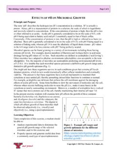
5 HE agar is particularly useful in the isolation of Shigella species. HE agar incorporates bile salts which allow for the SELECTIVE nature of this medium by inhibiting Gram-positive Figure 1. Stool culture on Hektoen enteric agar: bacteria , though these sales can also be Salmonella (black colonies) mixed with normal toxic for certain Gram-negative strains, fecal flora (orange-yellow colonies). Copyright 2020 by Chester R. Cooper, Jr. SELECTIVE and DIFFERENTIAL MEDIA , Page 2 of 7. but not Salmonella and Shigella species. HE agar also contains salicin, sucrose, and lactose as fermentable carbohydrates. Bromothymol blue and acid fuchsin in HE agar serve as acid-base indicators, whereas the presence of ferric ammonium citrate and sodium thiosulfate enable the detection of H2S, as noted by the formation of black-centered colonies.
6 After inoculation and incubation, colonies of Salmonella and Shigella species are green to bluish-green in color (Fig. 1). In addition, those Salmonella species that produce H2S shall appear as blue-green colonies with black centers. Alternatively, non-Salmonella and non- Shigella species that ferment lactose, sucrose, or salicin shall give rise to yellow-orange to salmon colored colonies. MacConkey Agar (MAC). MAC a SELECTIVE and DIFFERENTIAL medium for the isolation of Gram-negative bacilli, including coliforms and enteric pathogens. This medium was one of the earliest culture MEDIA for the cultivation and identification of enteric organisms and is also used in the isolation of pathogens from foods and coliforms in water samples.
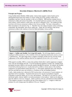
7 Like HE agar, MAC. incorporates bile salts, in addition to sodium chloride, which inhibits the growth of most Gram-positive bacteria . Differentiation of enteric microorganisms is achieved by the fermentation of lactose in the presence of bile salts and a neutral red indicator. Lactose-fermenting microbes form pink colonies surrounded by a zone of precipitated bile salts (Fig. 2). The production of acid from lactose Figure 2. Mixed fecal culture on MAC. fermentation changes the neutral red pH depicting a lactose fermenter (pink colonies) and indicator from colorless to red. The acid a non-lactose fermenter (white, clear colonies).
8 Production is also responsible for the formation of bile salt precipitation. Non-lactose-fermenters ( , Salmonella and Shigella species) develop as transparent, colorless colonies with no surrounding bile precipitate. Eosin Methylene Blue (EMB) Agar EMB agar was developed for the isolation of enteric bacilli. The incorporation of the dyes eosin and methylene blue inhibit the growth of Gram-positive bacteria in addition to serving as DIFFERENTIAL indicators for the fermentation of lactose. The production of acid from lactose fermentation results in an eosin-methylene blue dye complex being taken up by the microbial cell, thereby imparting a brown to blue-black colony appearance.
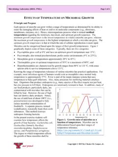
9 EMB agar has often been used to detect fecal and non-fecal coliforms and is recommended for use in the microbiological examination of potable water, waste water, dairy products, and foods. On EMB agar, isolated colonies of lactose-fermenting bacteria appear brown to blue-black in color. Escherichia coli grow as large, blue-black colonies that often possess a green metallic sheen (Fig. 3). However, not all E. coli strains produce a green metallic sheen. Hence, the green Copyright Chester R. Cooper, Jr. 2020. SELECTIVE and DIFFERENTIAL MEDIA , Page 3 of 7. metallic should not be used as the ultimate diagnostic for this microbe.
10 Non-lactose- fermenting colonies, such as Shigella and Salmonella species, appear transparent and colorless, though some strains of these two genera fail to grow on EMB agar. Occasionally, some Gram-positive bacteria , such as Enterococcus and Staphylococcus will grow on this medium, but usually as pinpoint colonies. Non- pathogenic, non-lactose-fermenting organisms may grow on EMB agar as well. Mannitol Salt Agar (MSA). Koch used MEDIA containing sodium chloride as a SELECTIVE agent for the isolation of Staphylococcus. Eventually, this observation was combined with the fact that Staphylococcus aureus usually Figure 3.