Transcription of SENSE ORGANS HANDOUT - soinc.org
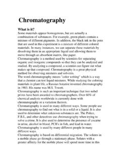
1 1 SENSE ORGANS HANDOUT Sensory Receptors - receive input, generate receptor potentials and with enough summation, generate action potentials in the neurons they are part of or synapse with 5 Types of Sensory Receptors - based on the type of stimuli they detect: 1. Mechanoreceptors - pressure receptors, stretch receptors, and specialized mechanoreceptors involved in movement and balance. 2. Thermoreceptors - skin and viscera, respond to both external and internal temperature 3. Pain receptors - stimulated by lack of O2, chemicals released from damaged cells and inflammatory cells 4. Chemoreceptors - detect changes in levels of O2, CO2, and H+ ions (pH) as well as chemicals that stimulate taste and smell receptors 5. Photoreceptors - stimulated by light Distribution of Receptors in the body.
2 Special senses mediated by relatively complex SENSE ORGANS of the head, innervated by cranial nerves vision, hearing, equilibrium, taste and smell General (somesthetic, somatosensory) receptors widely distributed in skin, muscles, tendons, joints, and viscera they detect touch, pressure, stretch, heat, cold and pain, blood pressure Special senses Sensation and perception Vision Eye Hearing Ear Equilibrium Ear Taste Taste receptors Smell Olfactory system General senses Skin Hot, cold, pressure, pain Muscles, joints, and tendons proprioceptors- stretch receptors respond to stretch or compression Pain Receptors somatic or visceral 2 SPECIAL senses Eye - Vision Processes Light energy is transduced into neural activity Neural activity is processed by the brain Note: For an analogy, you can imagine taking a picture with a camera.
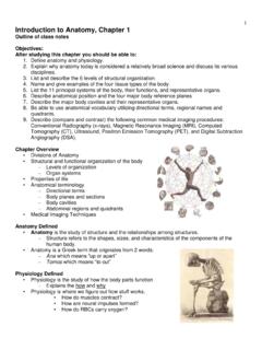
3 The eye is the camera, the retina, which is a specialized part of the brain at the back of the eye, is the film, and the parts of the brain that process visual information is the photoshop. Human visual systems permit light reflected off distant objects to be: Localized relative to the individual within his or her environment Identified based on size, shape, color, and past experience Perceived to be moving (or not) Detected in a wide variety of lighting conditions Sequence of events Light entering the eye is focused on the retina Retina converts light energy into neuronal activity Axons of the retinal neurons are bundled to form the optic nerves Visual information is distributed to several brain structures that perform different functions Eye the organ used to SENSE light Three layers 1.
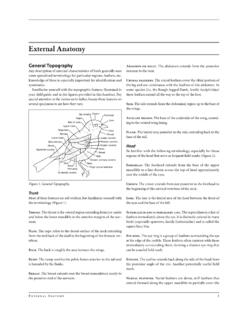
4 Outer layer consists of sclera and cornea 2. Middle layer consists of choroid, ciliary body and iris 3. Inner layer consists of retina Extraocular muscles--attached to the eye and skull and allow movement 3 anatomy of the Eye Gross anatomy Functions of the major parts of the eye: Sclera or Scleroid Layer (white of eye) the outermost layer that forms the eyeball- a tough protective layer of connective tissue that helps maintain the shape of the eye and provides an attachment for the muscles that move the eye Conjunctiva--membrane inside the eyelid attached to the sclera Cornea - the transparent surface covering the iris and pupil- a clear, dome-shaped part of the sclera covering the front of the eye through which light enters the eye Anterior Chamber a small chamber between the cornea and the pupil Aqueous Humor - fluid behind the cornea - the clear fluid that fills that anterior chamber of the eye and helps to maintain the shape of the cornea providing most of the nutrients for the lens and the cornea and involved in waste management in the front of the eye Choroid Layer - middle layer of the eye containing may blood vessels Ciliary Body - the ciliary body is a circular band of muscle that is connected and sits immediately behind the iris- produces aqueous humor, changes shape of lens for focusing.
5 And Iris - circular muscle that controls the diameter of the pupil - the pigmented front portion of the choroid layer and contains the blood vessels - it determines the eye color and it controls the amount of light that enters the eye by changing the size of the pupil (an albino only has the blood vessels not pigment so it appears red or pink because of the blood vessels) Lens - a crystalline structure located just behind the iris - it focuses light onto the retina Pupil - the opening in the center of the iris- it changes size as the amount of light changes (the more light, the smaller the hole) and it allows light to reach the retina Vitreous - a thick, transparent liquid that fills the center of the eye - it is mostly water and gives the eye its form and shape (also called the vitreous humor) Retina - axons of the retina leaving the eye - sensory tissue that lines the back of the eye.
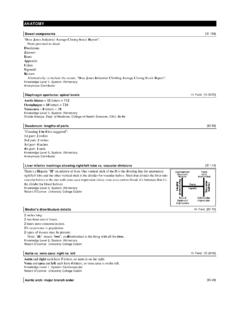
6 It contains millions of photoreceptors (rods for black & white and cones for color ) that convert light rays into electrical impulses that are relayed to the brain via the optic nerve Optic nerve - the nerve that transmits electrical impulses from the retina to the brain Opthalmoscopic appearance (Retina as seen through the pupil) Note: in photographs, the red appearance of the eye is actually the retina photographed. Double flash camera causes the pupil to constrict. Optic disk (blind spot)--no vision is possible o Blood vessels originate here. The vessels shadow the retina o Optic nerve fibers exit here o No photoreceptors Macula--area of the retina responsible for central vision (vs. peripheral) Fovea--center of the retina (where most of the cones are) 4 Common eye defects include myopia or nearsightedness where the eyeball is too long or the cornea is too steep; hyperopia or far sightedness where the eyeball is short or lens cannot become round enough: presbyopia where the muscles controlling the bulging of the lens become weak as we age; cataracts where the lens becomes fogged.
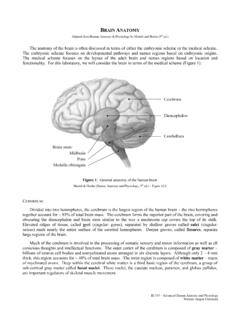
7 Nyctalopia or night blindness where vision is impaired in dim light and in the dark due to pigment rhodospin in the rods not functioning properly External features of the eye Cross sectional anatomy Lens--transparent surface that contributes to the formation of images (w/i 9 meters) Ciliary muscles--change the shape of the lens and allow focusing Vitreous humor--more viscous than the aqueous humor - Lies between the lens and the retina and provides spherical shape Retina - inner most layer of cells at the back of the eye - Transduces light energy into neural activity 5 Images the cornea and the lens help to produce the image on the retina images formed by the lens are upside down and backwards when they reach the retina two types of receptors on the retina Rods 125 million on a single retina extremely sensitive to all wavelengths of visible light but do not distinguish different color in dim light only rods are activated where one can see objects but not as sharp images and are not able to distinguish their color most dense in
8 Peripheral view nighttime vision Rods have a pigment called rhodospin As amount of light increases, the cones 7 million on a single retina mainly in central view are stimulated and the color becomes clear daytime vision There are three types of cones which distinguish the three colors blue, red, green Fovea point of central focus great density of cones - center of the eye's sharpest vision and the location of most color perception - the layers of the retina spread aside to let light fall directly on the cones Light stimulates rods and cones and sends impulse via optic nerve to brain areas for vision The Optic Nerve exits the eye just off center near the Fovea - the Optic Nerve exits is referred to as the Blind Spot due to the lack of the receptors in this area The two Optic Nerves come together at the Optic Chiasm located just under the hypothalamus - a crucial part of vision and perception must happen - cross-over of information from the right eye crosses over to the left side and visa versa happens here at the Optic Chiasm Information from each eye must be
9 Processed in both halves of the brain Information leaves the chiasm via the optic tract. Reorganized optic tract leaves the Optic Chiasm and passes onto the lateral geniculate nucleus At the lateral geniculate nuclei the information is separated, organized, and relayed to different areas of the visual cortex The different zones of the visual cortex process the different aspects of vision and information, taken from both visual fields, is processed and an image is perceived 6 EAR HEARING Outer Ear & ear canal brings sound into eardrum Eardrum vibrates to amplify sound & separates inner and middle ear Middle ear has 3 small bones or Ossicles = anvil, stirrup, stapes amplify sound (small bones) which vibrate sound Eustachian tube connects middle ear to throat and equalizes pressure on eardrum Cochlea in inner ear has receptors for sound & sends signals to brain via Auditory Nerve Process of hearing: Sound waves enter your outer ear and travel through your ear canal to the middle ear.
10 The ear canal channels the waves to your eardrum, a thin, sensitive membrane stretched tightly over the entrance to your middle ear. The waves cause your eardrum to vibrate. It passes these vibrations on to the hammer, one of three tiny bones in your ear. The hammer vibrating causes the anvil, the small bone touching the hammer, to vibrate. The anvil passes these vibrations to the stirrup, another small bone which touches the anvil. From the stirrup, the vibrations pass into the inner ear. The stirrup touches a liquid filled sack and the vibrations travel into the cochlea, which is shaped like a shell. Inside the cochlea, a vestibular system formed by three semicircular canals that are approximately at right angles to each other and which are responsible for the SENSE of balance and spatial orientation.