Transcription of Standard guide to fluorescence: instrument calibration and ...
1 NISTIR 7458 Standard guide to Fluorescence - instrument calibration and Validation Paul C. DeRose Biochemical Science Division National Institute of Standards and Technology Gaithersburg, MD 20899-8312 1 NISTIR 7458 Standard guide to Fluorescence - instrument calibration and Validation Paul C. DeRose Biochemical Science Division National Institute of Standards and Technology Gaithersburg, MD 20899-8312 October 2007 DEPARTMENT OF COMMERCE Carlos M. Gutierrez,Secretary TECHNOLOGY ADMINISTRATION Robert C. Cresanti,Under Secretary of Commerce for Technology NATIONAL INSTITUTE OF STANDARDS AND TECHNOLOGY James M.
2 Turner, Acting Director2 3 Table of Contents 1. 4 2. Wavelength 4 Low Pressure Atomic 5 Dy-YAG 5 Eu-doped glass or 5 Anthracene-doped PMMA .. 7 Ho2O3 solution or doped glass with diffuse reflector , scatterer or fluorescent dye .. 7 Xe Source 7 instrument Source with diffuse reflector or 9 Water 9 3. Spectral Slit Width 10 4. Linearity of the Detection 10 5. Spectral Correction of Detection System 11 Calibrated Light Source (CS) Tungsten 12 Calibrated Detector (CD) with Calibrated Diffuse reflector (CR).. 12 Certified Reference Materials (CRMs) .. 13 6. Spectral Correction of Excitation Beam 13 Calibrated Detector - Si Photodiode (CD Si).
3 14 Quantum 15 Si Photodiode (uncalibrated).. 15 7. calibration Curves for 15 Fluorophores with specified purity & 15 Fluorophore Solutions with specified concentration & 16 Molecules of Equivalent Soluble Fluorophore (MESF).. 16 8. Day-to-Day and instrument -to- instrument 16 Cuvette 17 Microwell Plate 17 Microarray 17 9. Limit of Detection and 17 10. 17 11. Fluorescence Quantum 18 Absolute 18 Optically Dilute Samples (A < ).. 18 Optically Dense 19 Integrating Sphere at 19 Relative 20 12. DEFINITION OF 20 13. Other Guideline/Recommendation 23 23 1. Scope To list the available materials and methods for each type of calibration or correction for fluorescence instruments (spectral emission correction, wavelength accuracy, etc.)
4 With a general description, the level of quality, precision and accuracy attainable, caveats, and useful references given for each entry. The listed materials and methods are intended for the qualification of fluorometers as part of complying with regulatory and other QA/QC requirements. Precision and accuracy or uncertainty are given at a 1 confidence level and are approximated in cases where these values have not been well 2. Wavelength Accuracy Methods for determining the accuracy of the emission (EM) or excitation (EX) wavelength for a fluorescence instrument are given here with an emphasis on monochromator (mono) based wavelength selection.
5 An example spectrum, which has been spectrally corrected, is given for each method. Table 1 : Summary of Methods for Determining Wavelength Accuracy NYNYNNYYE stablishedValueslimited range nmYY380nm-450nm (EM)310nm-380nm (EX)Anthracenein PMMA12limited range, calibration nmYY400nm-500nm (EX)Xe Source 12EX mono must be calibrated nmYMaybeUV-NIRXe Source + DR13EX mono must be calibrated nmYYUV-blueWater Ramanneed blankalignmentCaveats9-115, 631, 2 Ref. nmYMaybe330nm-800nm(EM or EX)Ho2O3 + DR nmYY570nm-700nm (EM)360nm-540nm (EX)Eu glass nmYY470nm-760nm (EM)255nm-480nm (EX)Dy-YAG crystal nm or betterYMaybeUV-NIR (EM)Pen Lamp UncertaintyOff-ShelfDrop-In regionSampleNYNYNNYYE stablishedValueslimited range nmYY380nm-450nm (EM)310nm-380nm (EX)Anthracenein PMMA12limited range, calibration nmYY400nm-500nm (EX)Xe Source 12EX mono must be calibrated nmYMaybeUV-NIRXe Source + DR13EX mono must be calibrated nmYYUV-blueWater Ramanneed blankalignmentCaveats9-115, 631, 2 Ref.
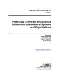
6 NmYMaybe330nm-800nm(EM or EX)Ho2O3 + DR nmYY570nm-700nm (EM)360nm-540nm (EX)Eu glass nmYY470nm-760nm (EM)255nm-480nm (EX)Dy-YAG crystal nm or betterYMaybeUV-NIR (EM)Pen Lamp UncertaintyOff-ShelfDrop-In regionSample 1 Certain commercial equipment, instruments, or materials are identified in this paper to foster understanding. Such identification does not imply recommendation or endorsement by the National Institute of Standards and Technology, nor does it imply that the materials or equipment identified are necessarily the best available for the purpose. 4 Low Pressure Atomic Lamps [1, 2] (nm)Relative IntensityThese low pressure atomic lamps, often referred to as pen lamps due to their size and shape, should be placed at the sample position and pointed toward the detection system for emission wavelength accuracy determination.
7 The emission wavelength selector ( EM-selector) is then scanned over the wavelength range of interest. (see Fig. 1) High accuracy is only achieved when the lamp is aligned properly into the wavelength selector, , the light fills the entrance slit of the monochromator. Atomic lines that are too close to other atomic lines to be resolved by the instrument should not be used, , may not be appropriate for low resolution instruments. Although these lamps can be placed at the excitation source position for excitation wavelength accuracy determination, weaker signals are typically observed, , by a reference detector, and alignment is more difficult than for the emission wavelength accuracy determination.
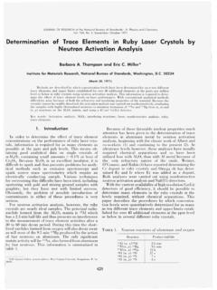
8 Fig. 1: Hg pen lamp spectrum Dy-YAG crystal [3] This sample is available in Standard cuvette format, so it can simply be dropped into a cuvette holder. An excitation or emission spectrum is then collected for an excitation or emission wavelength accuracy determination, respectively. (see Fig. 2) Peaks that are too close to other peaks to be resolved by the instrument should not be used, , may not be appropriate for low resolution instruments. Eu-doped glass [4] or PMMA [5, 6] This sample is available in Standard cuvette format, so it can simply be dropped into a cuvette holder.
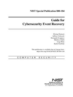
9 An excitation or emission spectrum is then collected for an excitation or emission wavelength accuracy determination, respectively. (see Fig. 3) Accurate peak 5 (nm)Relative Intensitypositions for this glass have not been well established and the positions of peaks can change somewhat depending on the particular glass matrix used. For these reasons, a one-time per sample determination of these peak positions using another wavelength calibration method is recommended. Fig. 2: Emission spectrum of a Dy-YAG crystal excited at nm. (nm)Relative Intensity Fig.
10 3: Emission spectrum of a Eu-ion-doped glass excited at 392 nm. 6 Anthracene-doped PMMA [7, 8] This sample is available in Standard cuvette format, so it can simply be dropped into a cuvette holder. An excitation or emission spectrum is then collected for an excitation or emission wavelength accuracy determination, respectively. (see Fig. 4) (nm)Relative Intensity Fig. 4: Emission spectrum of anthracene-doped PMMA excited at 360 nm. Ho2O3 solution or doped glass with diffuse reflector , scatterer or fluorescent dye [9-11] This sample is available in Standard cuvette format, so it can simply be dropped into a cuvette holder.