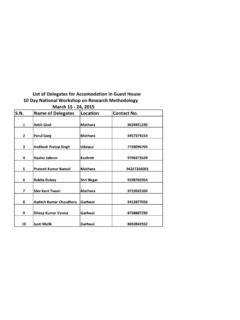
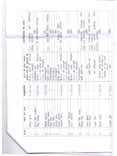
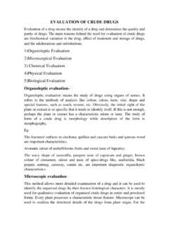
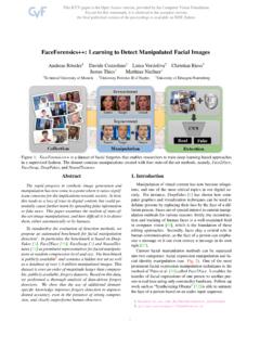
Transcription of Structure Of DNA & RNA - Jiwaji University
1 Structure Of DNA & RNA. By Himanshu Dev VMMC & SJH. DNA. DNA. Deoxyribonucleic acid DNA - a polymer of deoxyribo- nucleotides. Usually double stranded. And have double-helix Structure . found in chromosomes, mitochondria and chloroplasts. It acts as the genetic material in most of the organisms. Carries the genetic information A Few Key Events Led to the Discovery of the Structure of DNA. DNA as an acidic substance present in nucleus was first identified by Friedrich Meischer in 1868. He named it as Nuclein'. Friedrich Meischer In1953 , James Watson and Francis Crick, described a very simple but famous Double Helix model for the Structure of DNA.
2 FRANCIS CRICK AND JAMES WATSON. The scientific framework for their breakthrough was provided by other scientists including Linus Pauling Rosalind Franklin and Maurice Wilkins Erwin Chargaff Rosalind Franklin She worked in same laboratory as Maurice Wilkins. She study X-ray diffraction to study wet fibers of DNA. X-ray diffraction of wet DNA fibers The diffraction pattern is interpreted X Ray (using mathematical theory) Crystallography This can ultimately provide Rosalind information concerning the Structure Franklin's photo of the molecule She made marked advances in X-ray diffraction techniques with DNA.
3 The diffraction pattern she obtained suggested several structural features of DNA. Helical More than one strand 10 base pairs per complete turn Rosalind Franklin Maurice Wilkins DNA Structure DNA Structure is often divided into four different levels primary, secondary, tertiary and quaternary. DNA has three main components 1. Deoxyribose (a pentose sugar). 2. Base (there are four different ones). 3. Phosphate A, G, C or T A, G, C or U. O Base O Base O P O CH2 O P O CH2. 5 O 5 O. O 4 1 O 4 1 . H H H H. H H H H. Phosphate 3 2 . Phosphate 3 2 . OH H OH OH. Deoxyribose Ribose DNA Nucleotide RNA Nucleotide The Nitrogenous Bases THEY ARE DIVIDED INTO TWO GROUPS.
4 Pyrimidines and purines PYRIMIDINES (MADE OF ONE 6 MEMBER RING). Thymine Cytosine PURINES (MADE OF A 6 MEMBER RING, FUSED. TO A 5 MEMBER RING). Adenine Guanine THE RINGS ARE NOT ONLY MADE OF CARBON. Nitrogenous bases of DNA & RNA. Nucleotide Structure Nucleotides are formed by the condensation of a sugar, phosphate and one of the 4 bases The following illustration represents one nucleotide Phosphate Nitrogenous Bases Deoxyribose O Base O P O CH2. 5 O. O 4 1 . H H. H H. Phosphate 3 2 . OH H. Deoxyribose DNA nucleotide Base + sugar nucleoside Example Adenine + ribose = Adenosine Adenine + deoxyribose = Deoxyadenosine Base + sugar + phosphate(s) nucleotide Example Deoxyadenosine monophosphate (dAMP).
5 Deoxyadenosine diphosphate (dADP). Deoxyadenosine triphosphate (dATP). Deoxyadenosine triphosphate Doxyadenosine diphosphate Deoxyadenosine monophosphate Deoxyadenosine Adenine NH2. Phosphoester bond N. N. H. O O O N N. O P O P O P O CH2. 5 O. O O O 4 . H. 1 Base always H attached here H H. Phosphate groups 3 2 . HO H. Phosphates are attached here Deoxyribose Nucleotides are linked together by covalent bonds called phosphodiester linkage. A chemical bond that involves sharing a pair of electrons between atoms in a molecule. P. 5. 1. Base 4. Sugar 3 2. P. 5. 1 Base 4 Sugar 3 2. Backbone Bases O.
6 5 CH3. N. Thymine (T). H N O. O. O P O CH2. 5 O.. O 4 1 . H H. H H. 3 2 . NH2. H. N. N. Phosphodiester H Adenine (A). linkage O N N. O P O CH2. 5 O. O 4 1 . H H. H H. 3 2 NH2. H. H. N. Cytosine (C). O H N O. O P O CH2. 5 O.. O 4 1 . H H. H H Guanine (G). 3 2 . O. H. N H. N. H. O N N NH2. O P CH2 O. Single 5 O.. nucleotide O 4 1 . H H. Phosphate H H. 3 2 . OH H. Sugar (deoxyribose). 3 . DNA Double Helix & Hydrogen bonding Salient features of the Double-helix Structure of DNA: It is made of two polynucleotide chains, where the backbone is constituted by sugar-phosphate, and the bases project inside.
7 The two chains have anti- parallel polarity. It means, if one chain has the polarity 5' 3', the other has 3' 5'. 5' 3'. G C. T A. C G. A T. 3' 5'. DNA Double Helix & Hydrogen bonding The bases in two strands are paired through hydrogen bond (H-bonds). forming base pairs (bp). Adenine forms two hydrogen bonds with Thymine from opposite strand and vice-versa. Similarly, Guanine is bonded with Cytosine with three H-bonds. Based on the observation of Erwin Chargaff that for a double stranded DNA, the ratios between Adenine and Thymine; and Guanine and Cytosine are constant and equals one.
8 Hydrogen bond:-A chemical bond consisting of a hydrogen atom between two electronegative atoms ( , oxygen or nitrogen) with one side be a covalent bond and the other being an ionic bond. 3 Hydrogen bonds 2 Hydrogen bonds Erwin Chargaff's Experiment Chargaff pioneered many of biochemical technique for the isolation, purification and measurement of nucleic acids from living cells. It was known that DNA contained the four bases: A, G, C & T. Chargaff analyzed the base composition DNA isolated from many different species. THE HYPOTHESIS. An analysis of the base composition of DNA in different species may reveal important features about Structure of DNA.
9 Y Experimental level Conceptual level Solution of DNA +. 1. For each type of cell, extract the chromosomal proteins chromosomal material. This can be extract done in a variety of ways, including the use of high salt, detergent, or mild alkali treatment. Note: The chromosomes contain both DNA and protein. Protease DNA. 2. Remove the protein. This can be done in several ways, including treament with protease. Individual Acid bases 3. Hydrolyze the DNA to release the bases A T. from the DNA strands. A common way G C. A. to do this is by strong acid treatment. C. G T. C. G. 4. Separate the bases by chromatography.
10 AA A. Paper chromatography provides an easy A A A. way to separate the four types of bases. (The technique of chromatography is C. C C C CC C. described in the Appendix.). G G. G G GG G. 5. Extract bands from paper into solutions Origin and determine the amounts of each base T T. by spectroscopy. Each base will absorb TT T T. light at a particular wavelength. By examining the absorption profile of a sample of base, it is then possible to calculate the amount of the base. (Spectroscopy is described in the Appendix.). 6. Compare the base content in the DNA. from different organisms. The Data Interpretation of Data The compelling observation was that: Percentage of adenine=percentage of thymine Percentage of Cytosine=percentage of Guanine This observation became known as a Chargaff's Rule.