Transcription of Successful Function of Autologous iPSC-Derived Dopamine ...
1 Brief ReportSuccessful Function of Autologous iPSC-DerivedDopamine neurons following transplantation in aNon-Human Primate Model of Parkinson s DiseaseGraphical AbstractHighlightsdA non-human primate model tests cell transplantation for PDtherapydAutologous iPSC Dopamine neurons can provide long-termfunctional recoverydTransplanted cells survive for up to 2 years and reinnervatethe host brainAuthorsPenelope J. Hallett, Michela Deleidi, ..,Roger D. Spealman, Ole BriefA pre-clinical test of transplantation ofautologous iPSC- derived dopamineneurons in a cynomolgus monkey modelof Parkinson s disease provides proof ofprinciple for long-term innervation andfunctional benefit without a requirementfor et al., 2015, Cell Stem Cell16, 269 274 March 5, 2015 2015 Elsevier Stem CellBrief ReportSuccessful Function of Autologous iPSC-DerivedDopamine neurons following transplantation ina Non-Human Primate Model of Parkinson s DiseasePenelope J.
2 Hallett,1 Michela Deleidi,1 Arnar Astradsson,1 Gaynor A. Smith,1 Oliver Cooper,1 Teresia M. Osborn,1 Maria Sundberg,1 Michele A. Moore,1,2 Eduardo Perez-Torres,1 Anna-Liisa Brownell,1,3 James M. Schumacher,1 Roger D. Spealman,1,2and Ole Isacson1,4,*1 Neuroregeneration Research Institute, McLean Hospital and Harvard Medical School, Belmont, MA 02478, USA2 New England Primate Research Center, Harvard Medical School, Southborough, MA 01772, USA3 MGH/HST Athinoula A. Martinos Center for Biomedical Imaging, Massachusetts General Hospital and Harvard Medical School,Charlestown, MA 02129, USA4 Harvard Stem Cell Institute, Cambridge, MA 02138, transplantation of patient-specific in-duced pluripotent stem cell (iPSC)- derived neuronsis a potential clinical approach for treatment of neuro-logical disease. Preclinical demonstration of long-term efficacy, feasibility, and safety of iPSC-deriveddopamine neurons in non-human primate modelswill be an important step in clinical development ofcell therapy.
3 Here, we analyzed cynomolgus monkey(CM) iPSC- derived midbrain Dopamine neurons forup to 2 years following Autologous transplantationin a Parkinson s disease (PD) model. In one animal,with the most Successful protocol, we found thatunilateral engraftment of CM-iPSCs could provide agradual onset of functional motor improvementcontralateral to the side of Dopamine neuron trans-plantation, and increased motor activity, without aneed for immunosuppression. Postmortem analysesdemonstrated robust survival of midbrain-like dopa-minergic neurons and extensive outgrowth into thetransplanted putamen. Our proof of concept findingssupport further development of Autologous iPSC- derived cell transplantation for treatment of therapies offer an exciting opportunity to replace spe-cific populations of cells in neurodegenerative diseases wheresymptoms are defined by the loss of a specific cell type, suchas the degeneration of substantia nigra (SN) Dopamine neuronsin Parkinson s disease (PD).
4 The use of induced pluripotentstem cell (iPSC)- derived neurons as an Autologous cell sourceovercomes the current limitations posed by allogeneic donorcells in PD. Fetal ventral midbrain allografts can survive and func-tion in the human PD brain for over 18 years ( Freed et al.,2013, Soc. Neurosci., abstract;Hallett et al., 2014; Kefalopoulouet al., 2014; Mendez et al., 2005; Politis et al., 2010); however,such techniques will never become an easily accessible thera-peutic option for patients due to the requirement of fetal donortissue from elective abortions. Allografting in the brain also cre-ates a greater immune reaction over time compared with isoge-neic grafting (Duan et al., 1995; Morizane et al., 2013). The gen-eration of midbrain-like Dopamine neurons from patient-specificiPSCs and subsequent Autologous transplantation is a rationallong-term strategy for cell replacement in PD.
5 Previous reportsof Autologous transplantation in a non-human primate PD modelhave demonstrated the advantage of Autologous versus alloge-neic grafts and shown Dopamine neuron survival in the primatebrain for up to 6 months to 1 year after transplantation (Emborget al., 2013; Morizane et al., 2013; Sundberg et al., 2013). How-ever, the long-term Function , survival, and safety of iPSC-deriveddopamine neurons following Autologous transplantation in anon-human primate model of PD has not yet been studies were approved by the Harvard Medical SchoolInstitutional Animal Care and Use Committee (IACUC). To induceparkinsonism in cynomolgus monkeys (CMs), we administeredsystemic low-dose 1-methyl-4-phenyl-1,2,3,6-tetrahydropyri -dine (MPTP), which resulted in a progressive and persistentreduction in global motor activity and a stable bilateral parkinso-nian syndrome, including tremor, rigidity, bradykinesia, hypoki-nesia, posture/balance disturbances, and impairment in bothgross and fine motor skills (Table S1)(Brownell et al.)
6 , 1998a;Hantraye et al., 1992; Wu llner et al., 1994). All animals displayeda significant loss of Dopamine transporters (DATs) in the puta-men as measured by 11C-(2b-carbomethoxy-3b-(4-fluoro-phenyl ) tropane) (11C-CFT) binding potential, as previouslydescribed (Brownell et al., 1998a). In a successive series ofstudies, three MPTP-lesioned CMs (MF25-04, MF66-02, andMF27-04) received Autologous transplantation of CM-iPSC- derived neural cells into the putamen in order to assess thefunction and survival of engrafted Autologous iPSC-deriveddopamine neurons . CM-iPSCs from MF25-04 were differenti-ated using the protocol ofCooper et al. (2010), and CM-iPSCsfrom MF27-04 and MF66-02 were differentiated using the proto-col ofSundberg et al. (2013). No animals in this study receivedany immunosuppression for the duration of the recorded global daytime motor activity of MPTP-lesionedCMs that had received Autologous transplantation of iPSC- derived neural cells, using an automated activity monitor (Fig-ure 1A).
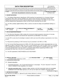
7 At 6 months after transplantation , daytime activitycounts in animal MF25-04 ( Autologous iPSC transplant) wereCell Stem Cell16, 269 274, March 5, 2015 2015 Elsevier by 146% compared with the stable parkinsonian pre- transplantation activity in this animal. Over the subsequent18 months of the study, motor activity in MF25-04 remainedelevated and ranged from 178% to 292% above pretransplanta-tion activity levels. At 2 years after transplantation , activity in thisanimal was 188% of stable MPTP baseline. The stable bilateralMPTP-lesion model used in this study provides an opportunityto assess asymmetry in movement functions following unilateraltransplantation, using movement analysis panel (MAP) testing(Figure 1B). A progressive improvement in the use of leftFigure 1. Functional Improvement of PD Motor Symptoms, Increased Dopamine Reuptake, and Reinnervation of the Transplanted Putamenafter Autologous transplantation of CM iPSC- derived Dopamine neurons (A) Differentiated CM-iPSCs were transplanted unilaterally into the putamen of three CMs with stable, bilateral parkinsonism (MF25-04, MF66-02, MF27-04).
8 Theanimals were followed for 1 2 years after transplantation . From 6 months after transplantation , functional improvement was observed in MF25-04, asdeterminedby a sustained increase in global daytime (6 to 6 ) activity. The non-transplanted control group and non-surviving transplant group represent the averagedata of n = 4 and n = 3 animals, respectively, and error bars show the SEM.(B) Fine-motor skills in MF25-04 were assessed using a computerized reaching task MAP. At 2 years after transplantation , MAP performance in the left upper limbwas significantly improved compared with pretransplantation values (p < , one-way ANOVA followed by Tukey s multiple comparison test). No changeinperformance was observed in the right upper limb. Data shown represent averages of five repeated tests (baseline), two repeated tests (1 year after trans-plantation), and three repeated tests (2 years after transplantation ).
9 Error bars represent the SEM.(C) Functional analysis of Dopamine reuptake in MF25-04 was measured by PET neuroimaging for11C-CFT, a marker of the DAT. Increased11C-CFT binding wasobserved in the transplanted putamen at 2 years after transplantation . White arrows indicate areas of hyperintense CFT PET signal.(D) Low-power photomicrograph of DAT immunostaining in the transplanted (right, R) and non-transplanted (left, L) putamen in MF25-04 shows reinnervation ofthe transplanted side. Deposits of grafted Dopamine neurons are indicated with boxes (g). IC, internal capsule; LGP, lateral globus pallidus; LV, lateral ventricle;3V, third ventricle; cc, corpus callosum.(E) Grafted Dopamine neurons were also labeled using tyrosine hydroxylase (TH). The boxed area is shown at higher magnification in the right. Robust survival ofdopamine neurons with outgrowth integration into the host putamen was Stem Cell16, 269 274, March 5, 2015 2015 Elsevier Inc.
10 (contralateral to the graft) upper limb motor Function , comparedwith baseline values, was observed by 12 months after trans-plantation in MF25-04, and this reduction reached significanceat 24 months after transplantation (p < , one-way ANOVA).Use of right upper limb motor Function in MF25-04 was not signif-icantly altered over the 24 months following the transplantationprocedure. The severity of parkinsonian signs was also ratedmonthly using a parkinsonian rating scale (Table S1). Weobserved a reduction in the hypokinesia subsection of theparkinsonian rating scale in MF25-04, from a score of 2 prior totransplantation (maximum possible score is 3) to a score of0 at 12 months after transplantation , and this remained stableat 0 until completion of the study (Table S1). Overall, the timecourse of functional recovery observed in MF25-04 was consis-tent with the developmental maturation, outgrowth, and connec-tivity of analogous fetal non-human primate Dopamine neurons (Redmond et al.)