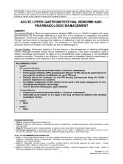
Transcription of SUMMARY RECOMMENDATIONS Level 1 a hemothorax.
1 DISCLAIMER: These guidelines were prepared by the Department of Surgical Education, Orlando Regional Medical Center. They are intended to serve as a general statement regarding appropriate patient care practices based upon the available medical literature and clinical expertise at the time of development. They should not be considered to be accepted protocol or policy, nor are intended to replace clinical judgment or dictate care of individual patients. EVIDENCE DEFINITIONS Class I: Prospective randomized controlled trial. Class II: Prospective clinical study or retrospective analysis of reliable data. Includes observational, cohort, prevalence, or case control studies.
2 Class III: Retrospective study. Includes database or registry reviews, large series of case reports, expert opinion. Technology assessment: A technology study which does not lend itself to classification in the above-mentioned format. Devices are evaluated in terms of their accuracy, reliability, therapeutic potential, or cost effectiveness. Level OF RECOMMENDATION DEFINITIONS Level 1: Convincingly justifiable based on available scientific information alone. Usually based on Class I data or strong Class II evidence if randomized testing is inappropriate. Conversely, low quality or contradictory Class I data may be insufficient to support a Level I recommendation.
3 Level 2: Reasonably justifiable based on available scientific evidence and strongly supported by expert opinion. Usually supported by Class II data or a preponderance of Class III evidence. Level 3: Supported by available data, but scientific evidence is lacking. Generally supported by Class III data. Useful for educational purposes and in guiding future clinical research. 1 Approved 04/27/ 2016 RETAINED hemothorax SUMMARY Retained hemothorax (RH) is complication of chest trauma that can lead to empyema, entrapped lung, and fibrothorax. When initial tube thoracostomy does not evacuate a hemothorax , options for management include a second tube thoracostomy, video-assisted thoracoscopy (VATS), or intrapleural fibrinolytic therapy.
4 Early VATS for retained hemothorax has been shown to decrease rates of empyema, decrease both intensive care unit and hospital days, and decrease the rate of conversion to thoracotomy. INTRODUCTION The definition of retained hemothorax varies throughout the literature. It is often defined as residual pleural blood >500ml in volume, blood occupying greater than one-third of the thoracic cavity, or any residual blood that cannot be drained after 72 hours of thoracotomy treatment (1). The incidence varies and can be as high as 20%, but in most studies is found to be 1-4% after initial tube thoracostomy for chest trauma (1,2). The most accepted complication of retained hemothorax is empyema.
5 DuBose et al. found that the overall incidence of empyema in retained hemothorax was 27% when defined by the presence of purulent pleural fluid, pleural fluid with a pH less than , or signs of infection or proven bacterial invasion of the pleural space on Gram stain or culture. Independent risk factors for the development of empyema included presence of rib fractures, Injury Severity Score >25, and need for additional therapeutic intervention. Patients with empyema had longer intensive care unit (ICU) and hospital stays, enforcing the need for prevention of empyema and other complications of retained RECOMMENDATIONS Level 1 VATS should be performed after initial chest tube thoracostomy has failed to evacuate a hemothorax .
6 VATS performed within 3-7 days has been shown to decrease complications of retained hemothorax . Level 2 There is no difference in hemothorax evacuation rate using a chest tube greater than 28 French. In patients with penetrating trauma, antibiotics with Gram positive coverage are effective at reducing infectious complications of tube thoracostomy including pneumonia and empyema. Chest computed tomography is the gold standard for diagnosing retained hemothorax . Level 3 Bedside ultrasound is superior to portable chest radiograph in diagnosing pleural fluid collections in blunt thoracic trauma. For patients who cannot undergo VATS, fibrinolytic therapy is an alternative for retained hemothorax .
7 2 Approved 04/27/ 2016 hemothorax (3). The literature suggests that VATS is an ideal way to evacuate blood from the pleural space and earlier rather than later intervention is beneficial in decreasing the morbidity of retained hemothorax (4,5). LITERATURE REVIEW Initial Chest Tube Management Chest tubes should be placed in all patients with hemothorax except those who meet criteria for operative intervention. The only prospective randomized controlled trial comparing chest tube size for traumatic hemothorax found no statistically significant differences in pain at site of insertion, efficacy of drainage, or rate of complications including retained hemothorax , need for additional tube drainage, or invasive procedures.
8 The chest tubes in this study were all placed non-emergently, and the sizes compared were 28-Fr to 32-Fr and 36-Fr to 40-Fr (6). A smaller study found a 50% decrease in insertion site pain and a trend toward lower analgesia requirements with 14-Fr pigtail drains compared to 28-Fr chest tubes; however, this paper was not powered to compare outcomes and the authors did not advocate for the use of 14-Fr pigtail drains for hemothorax (7). Currently, there is insufficient evidence to recommend one specific size or system over another for traumatic hemothorax ; however, there is no difference in the effectiveness or pain associated with chest tubes from 28-Fr to 40-Fr, and only chest tubes of 14-Fr or smaller have demonstrated statistically less pain at time of insertion, but these have not been evaluated for effectiveness in traumatic hemothorax .
9 Use of Antibiotics The use of antibiotics to reduce the risk of infectious complications of tube thoracostomy is controversial. The Eastern Association for the Surgery of Trauma (EAST) concludes that, there is insufficient published evidence to support any recommendation either for or against this practice (8). The Western Trauma Association concludes, it is not clear whether prophylactic antibiotics independently reduce the risk. Nevertheless, most centers administer .. antibiotics (9). The largest meta-analysis to date to answer this question includes 1,241chest tubes, making it far larger and comprehensive than the previous meta-analyses and largest individual randomized controlled trials.
10 This study found antibiotics with Gram-positive coverage reduced the overall infectious complication rate and lowered the risk of empyema. While this study was limited by heterogeneity in the antibiotics studied, the definitions of infectious complications, the types and sizes of chest tubes, and the method of placement, it is the most definitive publication on this topic to date (10). In patients with penetrating trauma, antibiotics with Gram-positive coverage are effective at reducing infectious complications of tube thoracostomy including pneumonia and empyema. Diagnosis of Retained hemothorax The gold standard imaging modality for diagnosing retained hemothorax is computed tomography (CT) of the chest, but alternative modalities have been studied.