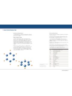
Transcription of T Cell Activation, In Vitro - Thermo Fisher Scientific
1 Bioassays BestProtocols Page 1 of 4 T Cell activation , In Vitro Research Use Only Revised 1-16-2013 Provided as a courtesy by eBioscience, An Affymetrix Company Copyright 2000-2013 eBioscience, Inc. Tel: or Fax: Introduction Mature T cells recognize and respond to the antigen/MHC complex through their antigen-specific receptors (TCR). The most immediate consequence of TCR activation is the initiation of signaling pathways including induction of specific protein tyrosine kinases (PTKs), breakdown of phosphatidylinositol 4,5-biphosphate (PIP2), activation of protein kinase C (PKC) and elevation of intracellular calcium ion concentration. These early events are transmitted to the nucleus and result in 1. clonal expansion of T cells 2. upregulation of activation markers on the cell surface 3. differentiation into effector cells 4. induction of cytotoxicity or cytokine secretion 5.
2 Induction of apoptosis One of the most common ways to assess T cell activation is to measure T cell proliferation upon in Vitro stimulation of T cells via antigen or agonistic antibodies to TCR. This protocol is written as a starting point for examining in Vitro proliferation of mouse splenic T- cells and human peripheral T cells stimulated via CD3. Critical parameters include cell density, antibody titer and activation kinetics. Protocol A: Stimulation of mouse peripheral T cells Materials 1X sterile PBS Anti-mouse CD3e, Clone 145-2C11 (Functional Grade, Cat. No. 16-0031, or Purified, Cat. No. 14-0031) Anti-mouse CD28, Clone (Functional Grade, Cat. No. 16-0281) Complete RPMI-1640 Sterile single-cell suspension of mouse spleen or lymph nodes 96-well flat-bottom microtiter plates with lids (Costar Cat. No. 3596) Concanavalin A, optional (ConA, Sigma Cat. No. C5275) Instruments Pipettes and pipettors, Multichannel pipettor Centrifuge 37 C, CO2 incubator Experiment Duration 2-18 hours to coat antibody to flask or plate 20 minutes preparation of spleen single cell suspension 20 minutes to set up the assay 2-4 days incubation Bioassays BestProtocols Page 2 of 4 T Cell activation , In Vitro Research Use Only Revised 1-16-2013 Provided as a courtesy by eBioscience, An Affymetrix Company Copyright 2000-2013 eBioscience, Inc.
3 Tel: or Fax: Experimental Procedure Step I: Antibody Coating of the Assay Plate Microwells: 1. Prepare a 5-10 g/mL solution of anti-CD3e (145-2C11) in sterile PBS. Calculate the number of wells required for each experimental condition and consider triplicate samples for each condition. For example, to coat one-half plate (48 wells) of antibody solution is required. Note: We have performed titration studies and found these concentrations of 145-2C11 to induce a maximal response. However, a pilot experiment to determine efficacy of other concentrations of this antibody to induce cellular activation can be performed. For costimulation studies using antibodies to other antigens, a suboptimal activation with anti-CD3 may be required. To achieve suboptimal activation via anti-CD3, a g/mL 145-2C11 antibody solution can be used. 2. Dispense 50 L of the antibody solution to each well of the 96-well assay plate.
4 For the control unstimulated wells, add 50 L of sterile PBS. 3. Tightly cover the plate with ParafilmTM to avoid sample evaporation and incubate at 37 C for 2 hours or prepare the plate one day in advance and keep at 4 C overnight. 4. Just before adding cells , remove the 50 L antibody solution with a multichannel pipettor. 5. Rinse each well with 200 L of sterile PBS and discard PBS. 6. Repeat step 5 to remove all unbound antibody from each well. Step II: Addition of cells : 1. Harvest spleen and prepare a single cell suspension under sterile conditions. Follow the BestProtocols: RBC Lysis of Mouse Splenocytes protocol to remove red cells . 2. Count cells and resuspend in complete RPMI-1640 at 106/mL. Note: This density is optimal for TCR-mediated T cell activation in our experiments. A titration of cell densities (2-3x10⁶ cells /mL to 105 cells /mL) is recommended for optimal activation in your studies.
5 3. After washing the wells with PBS (step 6 above), add 200 L of the cell suspension to each well and place in a humidified 37 C, 5% CO2 incubator. Note: For an additional stimulation control, incubate additional cells in 3 wells with Concanavalin A at 1-4 g/mL of culture medium. 4. Add soluble anti-CD28 to cells at 2 ug/mL. 5. Incubate for 2-4 days. Note: Proliferation of cells between days 2 and 4 gives a good proliferation response in our hands; however, this incubation time should be optimized by the end-user. 6. cells can be harvested and processed for your assay of interest. Buffer Recipes Complete RPMI-1640: 900 mL RPMI-1640 (Hyclone Cat. No. ) 100 mL FBS (Hyclone Cat. No. ) Heat inactivated (10% final) 1 mL 2-mercaptoethanol (Gibco BRL Cat. No. 21985-023) 10 mL L-Glutamine (Hyclone Cat. No. ) Antibiotic cocktail (optional) Bioassays BestProtocols Page 3 of 4 T Cell activation , In Vitro Research Use Only Revised 1-16-2013 Provided as a courtesy by eBioscience, An Affymetrix Company Copyright 2000-2013 eBioscience, Inc.
6 Tel: or Fax: Protocol B: Stimulation of human peripheral blood mononuclear cells Materials 1X sterile PBS Anti-human CD3: o Clone OKT3 (Functional Grade Cat. No. 16-0037) or Clone HIT3a (Functional Grade Cat. No. 16-0039) Anti-human CD28: o Clone (Functional Grade Cat. No. 16-0289) Complete RPMI-1640 Sterile pbmc 96-well flat-bottom microtiter plates with lids (Costar Cat. No. 3596) Instruments Pipettes and pipettors, Multichannel pipettor Centrifuge 37 C, CO2 incubator Experiment Duration 2-18 hours to coat antibody to flask or plate 30 minutes preparation of pbmc 20 minutes to set up the assay 2-4 days incubation Experimental Procedure Step I: Antibody Coating of the Assay Plate Microwells: 1. Prepare a 5-10 g/mL solution of anti-CD3e (OKT3 or HIT3a) in sterile PBS. Calculate the number of wells required for each experimental condition and consider triplicate samples for each condition.
7 For example, to coat one-half plate (48 wells) mL of antibody solution is required. Note: We recommend that you run a pilot experiment to determine efficacy of other concentrations of this antibody to induce cellular activation . For costimulation studies using antibodies to other antigens, a suboptimal activation with anti-CD3 may be required. To achieve suboptimal activation via anti-CD3, a g/mL OKT3 or HIT3a antibody solution can be used. 2. Dispense 50 L of the antibody solution to each well of the 96-well assay plate. For the control unstimulated wells, add 50 L of sterile PBS. 3. Tightly cover the plate with ParafilmTM to avoid sample evaporation and incubate at 37 C for 2 hours or prepare the plate one day in advance and keep at 4 C overnight. 4. Just before adding cells , remove the 50 L antibody solution with a multichannel pipettor. 5. Rinse each well with 200 L of sterile PBS and discard PBS.
8 6. Repeat step 5 to remove all unbound antibody from each well. Bioassays BestProtocols Page 4 of 4 T Cell activation , In Vitro Research Use Only Revised 1-16-2013 Provided as a courtesy by eBioscience, An Affymetrix Company Copyright 2000-2013 eBioscience, Inc. Tel: or Fax: Step II: Addition of cells : 1. Prepare pbmc and resuspend the cells at 1-2x10⁶/mL of complete RPMI. Note: This density of PBMCs is optimal for TCR-mediated T cell activation in our experiments. A titration of cell densities (2-3x10⁶ cells /mL to 105 cells /mL) is recommended for optimal activation in your studies. 2. After washing the wells with PBS (step 6 above), add 100 L of the cell suspension to each well. For each condition, use triplicate wells. 3. Add soluble anti-CD28 to cells at 2 ug/mL. 4. Place in a humidified 37 C, 5% CO2 incubator. 5. Incubate for 2-4 days. Note: Proliferation of cells between days 2 and 4 gives a good proliferation response in our hands; however, this incubation time should be optimized by the end-user.
9 6. cells can be harvested and processed for your assay of interest. Buffer Recipes Complete RPMI-1640: 900 mL RPMI-1640 (Hyclone Cat. No. ) 100 mL FBS (Hyclone Cat. No. ) Heat inactivated (10% final) 1 mL 2-mercaptoethanol (Gibco BRL Cat. No. 21985-023) 10 mL L-Glutamine (Hyclone Cat. No. ) Antibiotic cocktail (optional)