Transcription of The Accessory Ossicles of the Foot and Ankle; a …
1 THE JOURNAL OF ACADEMIC Review 106. EMERGENCY MEDICINE Derleme The Accessory Ossicles of the foot and Ankle; a Diagnostic Pitfall in Emergency Department in Context of foot and Ankle Trauma Ayak ve Ayak Bile i evresinde G r len Aksesuar Kemik ikler; Acil Serviste Ayak ve Ayak Bile i Travma Hastalar nda Tan sal Tuzak zkan K se Department of Orthoapedics and Traumatology, Antalya Education and Research Hospital, Antalya, Turkey Abstract zet Numerous normal anatomic variants including different Accessory Ossicles Ayak ve ayak bile i evresinde, normal anatomik varyant say lan ok say da and sesamoid bones are found around the foot and ankle region. They are aksesuar ve sesamoid kemik bulunmaktad r. Bunlar genellikle herhangi bir kli- usually of no clinical importance and are often recognized as an incidental nik nem ta mazlar ve tesad fi bir radyolojik bulgu olarak saptan rlar.
2 Ancak, radiographic finding. However, in the context of trauma, these Ossicles may travma varl nda, bu kemik ikler radyolojik olarak yanl tan ya ve gereksiz cause misdiagnosis due to various radiological pitfalls and this may result in tedavilere neden olabilirler. Bu sebeple, acil hekimlerinin normal anatomik under or overtreatment. Therefore, a thorough knowledge of these Accessory varyantlar tan mas , aksesuar ve sesamoid kemikler hakk nda bilgi sahibi Ossicles and sesamoid bones is important for the physician to recognize the olmas nemlidir. Bu derlemede aksesuar ve sesamoid kemikle ile kar kl a pathologic process and normal anatomic variants. This pictorial review sum- neden olabilecek k r klar ve ayak ve ayak bile i evresinde bulunan en yayg n marizes the most common Accessory Ossicles found around the foot and an- aksesuar kemik ikler zetlemi tir.
3 (JAEM 2012; 11: 106-14). kle in skeletally mature subjects and the fractures that may cause confusion Anahtar kelimeler: Aksesuar kemik, ayak, ayak bile i, k r k, os trigonum, os with Accessory Ossicles and sesamoid bones. (JAEM 2012; 11: 106-14) vesalianum os intermetatarseum, os supranaviculare, os supratalare, os pe- Key words: Accessory ossicle, foot , ankle, fracture, os trigonum, os vesalia- roneum, os subtibiale, os subfibulare, halluks sesamoid, os kalkaneus secun- num, os intermetatarseum, os supranaviculare, os supratalare, os peroneum, darius os subtibiale, os subfibulare, hallux sesamoids, os calcaneus secundarius Introduction Sesamoid bone is a small rounded bone embedded within a tendon or joint capsule.
4 Sesamoid bones are typically found in locations Trauma of the foot and ankle is commonly seen in patients pre- where a tendon passes over a joint. They prevent the friction senting to accident and emergency departments (1). Although, the between the tendon and the joint, protect the tendon and increase use of Ottawa Ankle Rules has reduced unnecessary radiographs, its biomechanical effect by changing the direction of pull of the ten- radiography still remains the most appropriate initial imaging modal- don. On the other hand, Accessory Ossicles are usually derived from ity in patients with suspected fractures (2). The decision to engage in the failure of union of secondary ossification centers to the main further imaging, indications for conservative treatment versus sur- bony mass.
5 They usually remain asymptomatic and are recognized as gery and consultation with an orthopedic surgeon or radiologist, an incidental radiographic finding (3). However, in the context of often rests on the emergency physician's interpretation of the radio- trauma, these Ossicles can be misdiagnosed as avulsion fractures, or graphical findings. Therefore, it is important for the emergency phy- a reverse situation in which an avulsion fracture can be evaluated as sician to be aware of abnormal and normal variants of bony lesions an Accessory ossicle is possible. of the foot and ankle and the various radiological pitfalls that may This pictorial review summarizes the most common Accessory cause confusion.
6 Many skeletal variations of the foot and ankle are Ossicles found around the foot and ankle and the fractures that may found, including different Accessory Ossicles and sesamoid bones. cause confusion with the Accessory Ossicles and sesamoid bones Correspondence to / Yaz ma Adresi: zkan K se, K lt r Mah. 3805. Sk. Durukent Sit. F BLok D: 22 Kepez, Antalya, Turkey Phone: +90 532 642 26 12 : Received / Geli Tarihi: Accepted / Kabul Tarihi: Copyright 2012 by Emergency Physicians Association of Turkey - Available on-line at Telif Hakk 2012 Acil T p Uzmanlar Derne i - Makale metnine web sayfas ndan ula labilir. zkan K se JAEM 2012; 11: 106-14 The Accessory Ossicles of the foot and Ankle 107. (Figure 1) (Table 1).
7 Their clinical significance, differential diagnosis, anatomy, epidemiology, related symptomatic disorders and the clinical and radiological features that help differentiate these distinct entities from fractures are also discussed and demonstrated. We aimed to summarize the necessary knowledge for emergency physi- cians to facilitate a correct and timely diagnosis and to decrease the rate of misdiagnosis that may lead to under or over-treatment, and unnecessary referrals. This review will focus only on the adult popu- lation because skeletally immature patients have different radiologi- cal characteristics due to the presence of physis or apophysis, and some Ossicles are radiologically evident at different periods of skel- etal maturation.
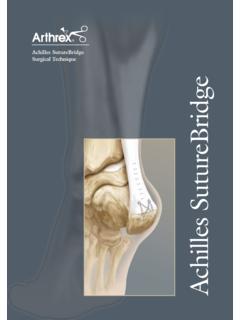
8 Os trigonum A secondary ossification center appears between the ages of 8. and 13 at the posterolateral aspect of the talus. Usually, this ossifica- tion center fuses with the talus within one year of its appearance (4). Fusion causes formation of a large posterolateral process which is referred to as a fused os trigonum', a Stieda's process', or a trigonal Figure 1. Locations of common Accessory Ossicles and sesamoid process'. When, it remains separate from the talus it is referred to as bones seen around the foot and ankle. 1. Accessory navicular bone 2. Os vesalianum 3. Os peroneum 4. Os intermetatarseum 5 Medial os trigonum (Figure 2) (5). The os trigonum articulates with the lat- hallux sesamoids 6 Lateral hallux sesamoid 7.
9 Hallux interphalangeal eral tubercle through a fibrocartilagenous synchondrosis (Figure 3). sesamoid bone 8. Metatarsophalangeal sesamoid bone 9. Os sub- The prevalence of the os trigonum ranges between 1-25% of the fibulare 10. Os subtibiale 11. Os trigonum 12. Os calcaneous population in different studies from Turkey and Japan (6-8). On con- secundarius 13. Os supratalare 14. Os supranaviculare Table 1. Summary of Accessory Ossicles and sesamoid bones that may simulate fractures around the foot and ankle Accessory /sesamoid bone Location Confused fractures Important clinical and radiographic findings and anatomical variants in acute avulsion fractures Os trigonum Posterolateral to the talus Fractures of posterior Positive nutcracker sign, remarkably sharp process of talus edges and discontinuity of the cortical lining Os vesalianum Adjacent to the base of 5th metatarsal base avulsion Tenderness over 5th metatarsal base, 5th metatarsal fracture, Jones fracture uncorticated sharp fracture line Os subfibulare Distal fibular tip Lateral malleolar avulsion Tenderness over distal fibular bony tip.
10 Fracture a missing part of the lateral malleolus, sharp uncorticated fracture line without sclerosis Os peroneum At the level of the Cuboid fractures, bipartite os Disruption in marginal cortical continuity of calcaneocuboid joint within peroneum cuboid, proximal migration and diastasis of the substance of the bipartite os peroneum peroneus longus tendon Os calcaneous secundarius Adjacent to the anterior Anterior superior calcaneus Tenderness over the anterior superior facet superior facet of calcaneus fracture of calcaneus, uncorticated often triangular fragment with sharp edges Os intermetatarseum Interspace between 1st and Lisfranc fracture-dislocation Disruption in tarsometatarsal joint alignment, 2nd metatarsal increased distance between cuneiforms Os subtibiale At the tip of medial malleolus Isolated medial malleolar Tenderness over the bony medial malleolar tip, avulsion fracture a missing part of the medial malleolus.