Transcription of The Cardiovascular System - Pearson
1 35611 When most people hear the term cardio-vascular System , they immediately think of the heart. We have all felt our own heart pound from time to time when we are ner-vous. The crucial importance of the heart has been recognized for ages. However, the Cardiovascular System is much more than just the heart, and from a scientific and medical standpoint, it is important to understand why this System is so vital to and day, minute after minute, our tril-lions of cells take up nutrients and excrete wastes.
2 Although the pace of these exchanges slows dur-ing sleep, they must go on continuously: when they stop, we die. Cells can make such exchanges only with the interstitial fluid in their immediate vicinity. Thus, some means of changing and refreshing these fluids is necessary to renew the nutrients and prevent pollution caused by the buildup of wastes. Like a bustling factory, the body must have a transportation System to carry its various cargoes back and forth. Instead of roads, railway tracks, and subways, the body s delivery routes are its hollow blood simply stated, the major function of the Cardiovascular System is transportation.
3 Using blood as the transport vehicle, the System carries oxygen, nutrients, cell wastes, hormones, and many other substances vital for body homeostasis to and from the cells. The force to move the blood The Cardiovascular SystemWHATHOWWHYThe Cardiovascular System delivers oxygen and nutrients to the body tissues and carries away wastes such as carbon dioxide via heart pumps blood throughout the body in blood vessels. Blood flow requires both the pumping action of the heart and changes in blood the Cardiovascular System cannot perform its functions, wastes build up in tissues.
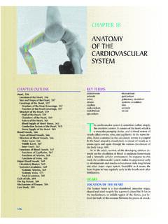
4 Body organs fail to function properly, and then, once oxygen becomes depleted, they will Building Vocabulary Coaching Activities for this chapter are assignable in Chapter 11: The Cardiovascular System 35711flanked on each side by the lungs (Figure ). Its pointed apex is directed toward the left hip and rests on the diaphragm, approximately at the level of the fifth intercostal space. (This is exactly where one would place a stethoscope to count the heart rate for an apical pulse.) Its broad posterosuperior aspect, or base, from which the great vessels of the body emerge, points toward the right shoulder and lies beneath the second and Walls of the HeartThe heart is enclosed by a sac called the pericar-dium (per i-kar de-um) that is made up of three layers: an outer fibrous layer and an inner serous membrane pair.
5 The loosely fitting superficial part of this sac is referred to as the fibrous pericar-dium. This fibrous layer helps protect the heart and anchors it to surrounding structures, such as the diaphragm and sternum. Deep to the fibrous pericardium is the slippery, two-layered serous pericardium. The parietal layer of the serous peri-cardium, or parietal pericardium, lines the inte-rior of the fibrous pericardium. At the superior aspect of the heart, this parietal layer attaches to the large arteries leaving the heart and then makes around the body is provided by the beating heart and by blood Cardiovascular System includes a muscular pump equipped with one-way valves and a System of large and small plumbing tubes within which the blood travels.
6 (We discussed blood, the sub-stance transported, in Chapter 10.) Here we will consider the heart (the pump) and the blood ves-sels (the plumbing ).The HeartAnatomy of the Heart Learning Objective Describe the location of the heart in the body, CPF|KFGPVKH[ KVU OCLQT CPCVQOKECN CTGCU QP CP appropriate model or , Location, and OrientationThe modest size and weight of the heart give few hints of its incredible strength. Approximately the size of a person s fist, the hollow, cone-shaped heart weighs less than a pound.]
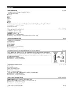
7 Snugly enclosed within the inferior mediastinum (me de-as-ti num), the medial section of the thoracic cavity, the heart is (a)Superiorvena cavaLeft lungAortaParietalpleura (cut)Pericardium(cut)PulmonarytrunkDiaph ragmApex ofheartFigure Location of the heart within the thorax. (a) Relationship of the JGCTV|CPF ITGCV XGUUGNU VQ VJG NWPIU (Figure continues on page 358.)358 Essentials of Human Anatomy and Physiologymyocardium is reinforced internally by a network of dense fibrous connective tissue called the skeleton of the heart.
8 The endocardium (en do-kar de-um) is a thin, glistening sheet of endothelium that lines the heart chambers. It is continuous with the linings of the blood vessels leaving and entering the heart. (Figure shows two views of the heart an exter-nal anterior view and a frontal section. As the ana-tomical areas of the heart are described in the next section, keep referring to Figure to locate each of the heart structures or regions.)Chambers and Associated Great Vessels Learning Objectives Trace the pathway of blood through the heart.
9 Compare the pulmonary and systemic heart has four hollow cavities, or chambers two atria (a tre-ah; singular atrium) and two ven-tricles (ven tr -kulz). Each of these chambers is lined with endocardium, which helps blood flow smoothly through the heart. The superior atria are primarily receiving chambers. As a rule, they are not important in the pumping activity of the heart. Instead, they assist with filling the ventricles. Blood flows into the atria under low pressure from the veins of the body and then continues on to fill the ventricles.
10 The inferior, thick-walled ventricles are the discharging chambers, or actual pumps of the heart. When they contract, blood is propelled out of the heart and into circulation. The right ven-tricle forms most of the heart s anterior surface; the left ventricle forms its apex (Figure ). The a U-turn and continues inferiorly over the heart surface. The visceral layer of the serous pericar-dium, or visceral pericardium, also called the epicardium, is part of the heart wall (Figure ). In other words, the epicardium is the innermost layer of the pericardium and the outermost layer of the heart wall.