Transcription of The Emerging Generation of Chromatography …
1 24 BioProcess International Oc tO b e r 2008 SupplementVACCINES PURIFICATIONThe Emerging Generation of Chromatography Tools for Virus Purification by Pete Gagnon Chromatography media and methods have evolved continuously since their introduction a half century ago. Traditional methods use columns packed with porous particles. They still dominate Chromatography applications in the field of virus purification, but the past 20 years have witnessed the ascendance of alternative supports, namely membranes and monoliths. These newer media exploit the familiar surface chemistries ion exchange, hydrophobic interaction, and affinity but they use unique architectures that offer compelling performance features.
2 Th E Ar C h I T E C Tu rE o f Ch r o mA To g rA p h y mE d I AA monolith can be defined as a continuous stationary phase cast as a homogeneous column in a single piece (1 3). Monoliths are further characterized by a highly interconnected network of channels with sizes ranging 1 5 m. The adsorptive surface is directly accessible to solutes as they pass through the column. The current Generation of preparative monoliths have bed heights ranging from a few millimeters to a few centimeters. Strictly speaking, membranes are monoliths. Their channel diameters similarly range from about to a few micrometers, but their bed heights are fractions of a millimeter. Traditional Chromatography media are prepared as porous particles and packed in columns.
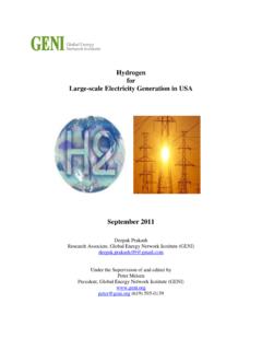
3 Most of their adsorptive surface area resides within shallow dead-end pores ranging 50 100 nm in size (4). Figure 1 compares the generalized structures of membranes, monoliths, and porous particles. mA S S Tr A N Sp o rTMembranes and monoliths differ most fundamentally from porous particle media in the mechanisms by which solutes are carried to and from their surfaces. The two primary modes of mass transport are diffusion and can be defined as the migration of solutes from an area of high concentration to an area of low concentration by incremental random thermal movement. Porous particle-based media rely almost exclusively on diffusive mass transport (4). Table 1 lists diffusion constants for representative solutes.
4 Two important points emerge: Diffusion is slow, and it becomes dramatically slower with increasing molecular size. As a direct consequence, dynamic binding capacity on porous particles decreases with increasing f low rates dramatically so for large solutes such as viral particles (1 7). Resolution also suffers, also in proportion to f low rate, and also more with large solutes. Diffusive limitations on porous particles can be compensated somewhat by reducing f low rate, but Figure 1: This comparison of generalized structures for stacked membranes, monoliths, and porous particles is not drawn to scale. Blue areas indicate structural material, white indicates areas of convective mass transport, and yellow indicates areas of diffusive mass transport.
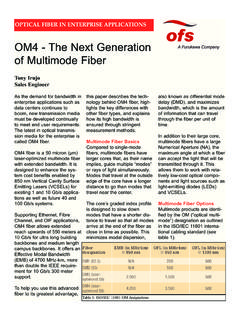
5 The entire volume of the particle is porous, but the pores are not highly interconnected, so only a small proportion of the particle is used (only diffusion-accessible pores are indicated). The white area between particles indicates void space. NFNCSBOF NPOPMJUI QPSPVT QBSUJDMFT26 BioProcess International Oc tO b e r 2008 Supplementthe diffusion constants of viral particles may be an order of magnitude slower than proteins, and few industrial users are willing to reduce f low rates to that degree (Table 1). Capacity and fractionation performance are thereby compromised. Pore exclusion is an additional limitation (8). Figure 2 illustrates a scale comparison of MVM and MuLV in relation to 100-nm diffusive pores and 1- m convective channels.
6 Pore size is roughly five times greater than the diameter of MVM, but MuLV is excluded and has access only to the external particle surface. This severely truncates binding capacity for large viral particles. Table 2 shows that many viral species have diameters above the exclusion limits of most porous particle-based media, but surface access within the monolith is unrestricted. Convection is movement imposed by an external force, in this case f luid f low delivered by the pumps in a Chromatography system. Convective mass transport is not limited by diffusion or by molecular size. A wine cork and a tree trunk both f low down a river at the same rate, which is determined by the velocity of the current.
7 The architecture of membranes and monoliths is designed specifically to take advantage of convective mass transport. Capacity and resolution are largely independent of f low rate, even at velocities 10 20 times faster than are commonly used with diffusive particles (1 3, 5 7). High f low rates offer numerous practical benefits. Process development and validation are accelerated, and manufacturing productivity is increased. Short process times may also be beneficial for live and attenuated viruses that are labile under the conditions used to conduct a particular purification step (8, 9). Figure 3 compares the dynamic binding capacity of a common particle-based anion exchanger with that of a monolithic anion exchanger.
8 DNA is used as a surrogate to illustrate the behavior of large solutes. Capacity on the monolith is 30 50 times higher (10), which allows for smaller columns and translates into proportional savings in media, buffer consumption, and use of expensive manufacturing space. Vo I d Co N Tr I b uT Io N SAnother key distinction between particles and monoliths is the presence or absence of an interparticle void volume. Fluid takes the path of least resistance through a bed of packed particles, that is, through the void volume rather than through the particles. That disfavors solute contact with Chromatography surfaces and contributes to lower virus binding capacity. Also, f luid friction at the particle surfaces causes formation of eddies (vortices that cause turbulent mixing, dilute peaks, and erode resolution).
9 Void space constitutes about 40% of total column volume, so it should be no surprise that eddy dispersion is a major cause of peak broadening in packed particle beds (7, 11, 12). As Figure 4 shows, eddies also create shear forces that can damage labile molecules (11 13). Eddy dispersion remains constant with increasing f low rate, but shear increases in direct proportion. Flow is laminar through monoliths; no eddies are formed (4, 7). In addition to minimizing shear, this assures an instantaneous response to changes in buffer composition, which maximizes Table 1: Diffusion constants for selected solutesSoluteSizeKdiffLight chain23 10 7 BSA66 10 7 IgG150 10 7 Urease480 10 7 IgM960 10 7 ETX2 10 7 CMV5 10 7 TMV40 10 10 10 9 ETX = endotoxin; CMV = Cucumber mosaic virus; TMV = Tobacco mosaic virusTable 2: Approximate diameters of selected viral particlesVirusDiameterAAV20 26 nmMVM25 nmRhinovirus30 nmHBV42 nmAdenovirus59 67 nmEBV80 100 nmHIV100 120 nmHSV110 200 nmMuLV120 150 nmAAV = adenoassociated virus; MVM = minute virus of mice.
10 HBV = hepatitis B virus; EBV = Epstein-Barr virus; HIV = human immunodeficiency virus; HSV = herpes simplex virus; MuLV = murine leukemia virusFigure 2: In this scale comparison of particle pores and monolith channels, illustrated pore size is 100 nm, and channel size is 1 m (1,000 nm). Murine leukemia virus (MuLV, 150 nm diameter ), minute virus of mice (MVM, 25 nm diameter), and immunoglobulin G (IgG, 12 nm hydrodynamic diameter) are added for reference. Blue areas indicate structural material, white areas indicate areas of convective mass transport, yellow indicates areas of diffusive mass transport, and arrows mark the direction of flow. As indicated, MuLV cannot enter the pores and has access only to the exterior of the particle.