Transcription of THE KNEE IN CEREBRAL PALSY: CURRENT MANAGEMENT …
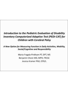
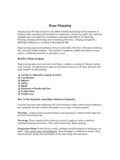
1 1 THE knee IN CEREBRAL palsy : CURRENT MANAGEMENT FROM LESSONS LEARNT through three dimensional GAIT ANALYSIS[1] H Kerr Graham1,2,3, Paulo Selber2, Jill Rodda1, Jeffrey Young2 1 Murdoch Childrens Research Institute, Royal Children s Hospital, Melbourne, Australia. 2 The Department of Orthopaedics, Royal Children s Hospital, Melbourne, Australia, 3 The University of Melbourne, Melbourne, Australia. INTRODUCTION Orthopaedic priorities by topographic classification Hemiplegia: Foot/Ankle: equinus, equinovarus, equinovalgus. Diplegia: knee : sagittal plane balance for walking. Quadriplegia: Spine/Pelvis/Hip: sitting balance THE EVALUATION Diagnostic Matrix Davids et al [2] information is gathered from the following five sources (Figure 1): 1.
2 Clinical history: GMFCS, FMS, FAQ 2. Physical examination 3. Instrumented gait analysis 4. Special investigations including radiology 5. Examination under anesthesia Figure 1: Diagnostic Matrix 2 Physical Examination Of The knee Muscle spasticity (R1 vs. R2) Muscle Contracture knee Joint Contracture Popliteal angle Duncan Ely/Prone rectus Fixed Flexion Deformity Gait Analysis Understanding the gait pattern and the interrelationship of the hip, knee , and ankle helps guide treatment 4 Sagittal knee Patterns: Sutherland and Davids[3]: 1) jump knee increased knee flexion in early stance.
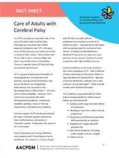
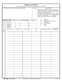
3 Normal knee extension in late stance 2) Crouch increased knee flexion and ankle dorsiflexion in stance 3) Stiff knee decreased knee flexion in swing 4) Recurvatum knee excessive knee extension in stance 4 Sagittal Gait Patterns: Rodda and Graham[4] (Figure 2): 1) True equinus hip normal; knee normal; ankle equinus 2) jump Gait hip normal; knee flexed; ankle equinus 3) Apparent Equinus hip/ knee flexed; ankle plantigrade 4) Crouch Gait hip/ knee flexed; ankle dorsiflexed Figure 2: Four sagittal gait patterns in spastic diplegia Rodda and Graham[4]. 3 knee INTERVENTIONS IN SPASTIC DIPLEGIA Non Operative MANAGEMENT Strengthening: Important component of MANAGEMENT particularly o Post multilevel orthopaedic surgery (and even in preparation) o Mild cases of diplegia o Adolescence Short term: strength improves with isometric training[5, 6] Detraining occurs as early as six weeks after cessation of the program[6] The relationship between improved strength and gait is uncertain.
4 Gait kinematics show wide variability following an eight week progressive resistance exercise program[7] Spasticity MANAGEMENT For knee Dysfunction[8, 9]: Oral medications diazepam (Valium) for acute postoperative pain and spasm MANAGEMENT . Not for chronic MANAGEMENT . Botulinum Toxin A (BoNT A) targets specific muscle groups; o Hamstring injections Corry et al[10] Improves knee extension at initial contact six degrees Maximal knee extension in stance eight degrees Improvements no longer present at 12 weeks o defers multilevel orthopaedic surgery until an appropriate age Molenaers et al[11] o effective in the short term, but is never definitive Selective dorsal rhizotomy useful for severe hypertonia in carefully selected cases Intrathecal baclofen pump for generalized hypertonia Operative MANAGEMENT Flexed knee gait cannot be managed by a single approach.
5 Applying the diagnostic matrix (clinical history, physical examination, gait analysis, radiology, and examination under anesthesia) helps determine the surgical dose. Distal Hamstring Lengthening Establishing whether the hamstrings are or are not short and contracted is paramount prior to undertaking any hamstring lengthening procedure as the prevalence of short, contracted hamstrings in spastic diplegia has been shown by studies using muscle length modeling, to be less frequent than previously thought[12]. A variety of techniques for hamstring lengthening are utilized: Semimembranosus lengthening by a number of circular stripes in the fascia over the distal muscle, followed by gentle extension of the knee , allows lengthening in continuity while preserving the underlying muscle and muscle function.
6 Gracilis and semitendinosus lengthening by simple tenotomy, Z lengthening or intramuscular lengthening have been described. 4 Lateral hamstring lengthening in addition to medial hamstring lengthening is more controversial due to: Risk of knee recurvatum [13]. Risk if increased anterior pelvic tilt[14]. Presenters Preferred Method Less commonly performing isolated distal hamstring lengthening because the knee dysfunction is often too mild or too severe to benefit. We lengthen the medial hamstrings only, to minimize the risk of recurvatum and minimize the risk of increased anterior pelvic tilt.
7 To maintain the integrity of the muscle tendon unit, the semimembranosus is fractionally lengthened with one or two circumferential stripes in the fascia, and the semitendinosus and gracilis are lengthened by intramuscular technique, similar to that described for the lengthening of the tibialis posterior[15] (figure 3B). We always use intramuscular tenotomy to preserve continuity and function. Now we almost never lengthen biceps femoris. Distal hamstring lengthening is combined with other procedures, such as a semitendinosus transfer, for mild to moderate degrees of fixed flexion deformity at the knee , greater than 5 degrees.
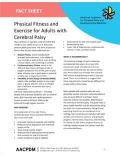
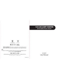
8 5 Figure 3: Schematic representation of knee interventions in spastic diplegia. A) For spastic muscle contracture, spasticity MANAGEMENT may include botulinum toxin A injections (BoNT A), selective dorsal rhizotomy (SDR) and occasionally intrathecal baclofen (ITB). B) Fixed muscle contractures are addressed by medial distal hamstring lengthening. C,D) Useful tendon transfers include a rectus femoris transfer or semitendinosus transfer. E) Fixed knee flexion contracture may be addressed by supracondylar extension osteotomy and patellar tendon shortening. F) When growth is remaining, guided growth can be employed, applying 8 plates across the anterior distal femoral physis.
9 6 Rectus Femoris Transfer Distal hamstring lengthening in isolation improves knee extension during stance phase but results in decreased flexion during swing phase and increased stiffness[16]. This may result in clearance problems, toe scuffing and increased energy expenditure[17]. A solution to this problem is to combine medial hamstring lengthening with transfer of the rectus femoris. The indications include: GMFCS level the best results of rectus femoris transfer are in GMFCS I and II patients[18]. Rectus femoris spasticity confirmed by the Duncan Ely or prone rectus test [17, 19].
10 Kinematic variables including decreased peak knee flexion in swing phase[19], a decreased knee range of motion during swing phase[17, 20], a decreased overall knee range of motion during the gait cycle, and a delay in the timing of peak knee flexion[19]. Dynamic EMG showing prolonged rectus firing during swing phase [17, 20]. With respect to surgical technique: Distal rectus femoris transfer yields better results than either proximal or distal rectus femoris lengthening [20 22]. No evidence that one site of transfer is superior to another [23, 24]. Medially, the most commonly used recipient tendons for the rectus femoris transfer are the semitendinosus, the gracilis or the sartorius.