Transcription of The management of cardiac arrest - ALSG
1 47 CHAPTER 6 The management of cardiac arrest LEARNING OBJECTIVES In this chapter you will learn: How to assess the cardiac arrest rhythm and perform advanced life support INTRODUCTION cardiac arrest has occurred when there is no effective cardiac output. Before any specific therapy is started effective basic life support must be established as described in chapter 4. Four cardiac arrest rhythms will be discussed in this chapter: 1. Asystole 2. Pulseless electrical activity (including electro mechanical dissociation) 3. Ventricular fibrillation 4 Pulseless ventricular tachycardia THE management OF cardiac arrest 48 The four are divided into two groups: two that do not require defibrillation (called non-shockable ) and two that do require defibrillation ( shockable ).
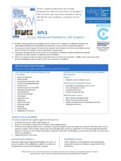
2 The cardiac arrest algorithm is shown in Figure Stimulate andassess responseOpen airwayCheck breathing5 rescue breathsCheck pulseCheck for signs of circulationCPR15 chest compressions2 ventilationsAssessrhythmAsystole andPEAVF/VTVentilate with highconcentration O2 Adrenaline10 mcg/kg IV or IOContinue CPRI ntubateIV/IO access4 min CPRDC Shock 4J/kgDC Shock 4J/kgAdrenaline thenDC Shock 4J/kgAmiodarone then DC Shock 4J/kgAdrenaline thenDC Shock 4J/kgDC Shock 4J/kg 2 min CPR,check monitor 2 min CPR,check monitor 2 min CPR,check monitor 2 min CPR,check monitor 2 min CPR,check monitorIntubate, High flow O2IV/IO accessConsider4 HsHypoxiaHypovolaemiaHyper/hypokalaemia / MetabolicHypothermia4 TsTension pneumothoraxCardiac TamponadeToxic substancesThromboembolic phenomenaConsider alkalising agents2 min CPR,check monitorAdrenaline dose 10 mcg/kgAmiodarone dose 5mg/kgCheck monitorevery 2 minutesIntubateIV/IO accessShockableNonshockable Figure cardiac arrest algorithm NON SHOCKABLE RHYTHMS This includes asystole and pulseless electrical activity.
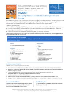
3 THE management OF cardiac arrest 49 Asystole This is the most common arrest rhythm in children, because the response of the young heart to prolonged severe hypoxia and acidosis is progressive bradycardia leading to asystole. The ECG will distinguish asystole from ventricular fibrillation, ventricular tachycardia and pulseless electrical activity. The ECG appearance of ventricular asystole is an almost straight line; occasionally P-waves are seen. Check that the appearance is not caused by an artifact, a loose wire or disconnected electrode.
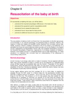
4 Turn up the gain on the ECG monitor. Figure Asystole Pulseless Electrical Activity (PEA) This is the absence of a palpable pulse or other signs of circulation despite the presence on the ECG monitor of recognisable complexes which normally produce a pulse. PEA is treated in the same way as asystole and is often a pre-asystolic state. Pulseless electrical activity may be due to an identifiable and reversible cause. In children, trauma is most often associated with a reversible cause of PEA. This may be severe hypovolaemia, tension pneumothorax or pericardial tamponade.
5 PEA is also seen in hypothermic patients and in patients with electrolyte abnormalities, including hypocalcaemia from calcium channel blocker overdose. Rarely in children it may be seen after massive pulmonary thromboembolus. Figure Pulseless Electrical Activity (PEA) management of Asystole/PEA The first essential is to establish ventilations and chest compressions effectively. Ventilations are provided initially by bag and mask with high concentration oxygen. Ensure a patent airway, initially using an airway manoeuvre to open the airway and stabilising it with an airway adjunct.
6 THE management OF cardiac arrest 50 Provide effective chest compressions at a rate of 100 per minute with a compression/ ventilation ratio of 15: 2. The child should have a cardiac monitor attached and the heart s rhythm assessed. Although the procedures to stabilise the airway and gain circulatory access are now described sequentially, they should be undertaken simultaneously under the direction of a resuscitation team leader. If asystole or PEA is identified give adrenaline (epinephrine) 10 micrograms per kilogram intravenously or intraosseously.
7 Adrenaline (epinephrine) is the first line drug for asystole. Through -adrenergic mediated vasoconstriction, its action is to increase aortic diastolic pressure during chest compressions and thus coronary perfusion pressure and the delivery of oxygenated blood to the heart. It also enhances the contractile state of the heart and stimulates spontaneous contractions, The intravenous or interosseous dose is 10 micrograms/kg ( ml of 1:10,000 solution). This is best given through a central line but if one is not in place it may be given through a peripheral line.
8 In the child with no existing intravenous access the intraosseous route is recommended as the route of choice as it is rapid and effective. In each case the adrenaline (epinephrine) is followed by a normal saline flush (2-5 mls). If circulatory access cannot be gained, the tracheal tube can be used but this is the least satisfactory route and should be avoided if possible as it may cause transient adrenergic effects, decreasing coronary perfusion. If the route is used, ten times the intravenous dose (100 micrograms/kg) should be given.
9 The drug should be injected quickly down a narrow bore suction catheter beyond the tracheal end of the tube and then flushed in with 1 or 2 mls of normal saline. As soon as is feasible a skilled and experienced operator should intubate the child s airway. This will both control and protect the airway and enable chest compressions to be given continuously, thus improving coronary perfusion. Once the child has been intubated and compressions are uninterrupted, the ventilation rate should be 10 per minute. It is important for the team leader to assess that the ventilations remain adequate when chest compressions are continuous.
10 The protocol for asystole and PEA is shown in Figure Figure Protocol for asystole and PEA During and following the administration of adrenaline (epinephrine), chest compressions and ventilations should continue. Giving chest compressions is tiring for the operator so the team leader should continuously ensure that the compressions are adequate and change the operator every few minutes. THE management OF cardiac arrest 51 At intervals of about 2 minutes briefly pause in the delivery of chest compressions to assess the rhythm on the monitor.