Transcription of The Muscular System PDF - Class Videos for …
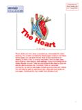
1 11:10 am, Feb 25, 2008 Entire PDF. Audio File In Real Media Format The Muscular System Jim Swan 1. These slides are from Class presentations, reformatted for static viewing. The content contained in these pages is also in the Class Notes pages in a narrative format. Best screen resolution for viewing is 1024 x 768. To change resolution click on start, then control panel, then display, then settings. If you are viewing this in Adobe Reader version 7 and are connected to the internet you will also be able to access the enriched links to notes and comments, as well as web pages including animations and Videos .
2 You will also be able to make your own notes and comments on the pages. Download the free reader from [ ]. 1. 3 Types of Muscle Tissue Muscle Type Location Characteristics Control Attached to the Long, cynlindrical Skeletal cells; Voluntary bones for multinucleated, movement striated Short, branching cells, Involuntary Cardiac Muscle of the mononucleated, faintly Heart striated. Forms myogenic functional syncytia. Smooth Single Unit: GI, Muscle Respiratory, & Small oblong cells, Involuntary Genitourinary tract mononucleated, myogenic mucous membranes. also may form a Multi-unit: smooth functional muscle in blood syncytium.
3 2. vessel walls. 2. Skeletal Muscle nuclei Connective endomysium separates cells. Striations =. dark bands Myofibrils fill sarcoplasm 3. The nuclei and other organelles of skeletal muscle cells are found next to the sarcolemms and the majority of the sarcoplasm is filled with the contractile machinery of the cell, the myofibrils. Skeletal muscle cells are derived from individual myocytes which fuse to produce a mature multinucleated muscle fiber. There are few if any of the precursor myocytes found in a mature muscle, and so muscles produce no new cells after maturity. Individual cells respond to training by enlarging and building myofibrils and other components.
4 3. Skeletal Muscle photomicrographs Dark striations = A-bands, the light areas between are the I-bands. Z-line. From one Z. line to the next is a sarcomere. Z-line The sarcolemma is the cell membrane 4. 4. Cardiac Muscle Intercalated disks Faint striations Branching cells connect to form network. mononucleated The action potential travels through all cells connected together in a syncytium causing them to function as a unit. 5. Cardiac muscle cells are much shorter than cells in skeletal muscle and they branch to connect to neighboring cells through specialized membranes called intercalated disks to form a network called a syncytium.
5 5. Smooth Muscle nucleus Spindle -shaped mononucleated smooth muscle cell. 6. Smooth muscle cells connect to form single-unit syncytia similar to cardiac muscle. But impulses and contractions occur much more slowly in smooth than in cardiac muscle. 6. Smooth Muscle Arrangement In the intestine smooth muscle forms two distinct layers, one running along, the other running around the organ. Together these layers cause movements which propel the contents. The circular layer runs around the intestine and its contraction causes segmentation The longitudinal layer runs along the 7. intestine; it causes wave-like contractions.
6 7. Types of Smooth Muscle Fibers Single unit smooth muscle cells connected to function as a single unit (syncytium). in GI tract Multiunit smooth muscle cells grouped into many contractile units controlled by the nervous System . in blood vessel walls and sphincters in GI tract. 8. 8. Structure of a Skeletal Muscle Tendon attachment Belly contains cells epimysium Fibrous covering perimysium fascicle { Surrounds fascicle Cell (fiber). endomysium Figure Surrounds cells (fibers). 9. The hierarchy of connective tissues associated with a skeletal muscle provide a continuous connection between muscle cells and their action on a bone or other attachment.}
7 At the same time cells are effectively separated from one another and each is controlled by a separate nerve fiber. 9. Functional Characteristics of Skeletal Muscle Excitability (responsiveness) muscles can be stimulated by electrical, chemical, and physical means. Contractility a muscle responds to stimuli by contracting. Elasticity muscles tend to recoil to their resting length. Extensibility muscles can be stretched beyond their resting length. 11. 11. Muscle Attachments Tendons attach muscle to bone. Aponeuroses broad, flat, tendinous attachment. Origin more fixed point of attachment. Insertion more movable point of attachment.
8 Muscle action pulls insertion toward the origin. A muscle can only pull, it cannot push. 10. 10. Types of Muscle Contractions Agonist the prime mover; the muscle which performs the movement in question. Antagonist the muscle that performs the opposing movement to that of the agonist. Both muscles contract (exert tension) regardless of which is the agonist or antagonist. On-center movement that of the agonist Off-center (eccentric) movement that of the antagonist 12. The antagonist may actually be stretching while it is generating tension (contracting). 12. Knee Extension Agonist: the rectus femoris (quadriceps femoris).
9 Antagonist: the biceps femoris (hamstrings). 13. 13. Knee Flexion Agonist: the biceps femoris (hamstrings). Antagonist: the rectus femoris (quadriceps femoris). 14. 14. Synergists muscles which work together to perform a movement; often differs from the movement either performs when working alone. Fixators muscles which work to keep a part from moving; stabilizers, neutralizers. Assignment: make a list of 5 each antagonists, synergists, and fixators. 15. For each example you will need a couple of muscles: for antagonists you will need the two muscles which are antagonistic, for synergists the two or more muscles which work together, and for fixators the part of the body which is fixed and under what conditions.
10 15. A Muscle Cell nucleus = A Fiber myofibril striations sarcolemma {. I Band Striation = A Band Titin proteins A sarcomere actin myosin 16. Look at the video clip showing muscle hierarchy. From the largest to smallest the hierarchy is as follows: whole muscle is composed of bundles of cells called fasciculi, individual cells are composed of myofibrils, which are organized of myofilaments of actin and myosin and other proteins arranged in a specific way. 16. The Sarcomere A-Band I-Band Z-disk H-zone Titin {. Actin filaments Myosin filaments (thin bands) (thick bands). 17. Titin proteins are part of the structural support for the myofilaments of the sarcomere and also a part of the series elastic elements along with other proteins and connective tissues.}}