Transcription of The Umbilical Cord - Yale School of Medicine
1 Sunday, October 29, 2006 Page 1 of 14 The Umbilical Cord (from The Encyclopedia of Reproduction) Harvey J. Kliman, , yale University School of Medicine I. Introduction II. Formation and structure of the Umbilical cord III. Abnormal Umbilical cord development IV. Pathologic processes affecting the Umbilical cord V. Umbilical cord length and twisting VI. Umbilical cord insertion VII. Diagnostic utility of the Umbilical cord Glossary Allantois primitive excretory duct Cord prolapse passage of the Umbilical cord through the cervix prior to delivery of the infant Funisitis inflammatory cell infiltrate in the Umbilical vessels walls and Wharton s jelly Insertion point at which the Umbilical cord attaches to the placenta Meckel s diverticulum persistent outpouching of bowel contents through the abdominal wall at the umbilicus Omphalomesenteric duct remnant remains of the yolk sac stalk within the proximal portion of the Umbilical cord Vasa previa Umbilical cord vessels, usually in a case of a velamentous insertion, which are overlying the internal cervical os Velamentous insertion of the Umbilical cord into the external membranes The Umbilical Cord Harvey Kliman Sunday.
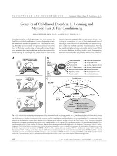
2 October 29, 2006 Page 2 of 14 Wharton s jelly proteoglycan rich matrix in which the Umbilical vessels are embedded Yolk sac outpouching of the endoderm which serves as the site of initial blood cell formation The Umbilical Cord Harvey Kliman Sunday, October 29, 2006 Page 3 of 14 I. Introduction The Umbilical cord is the lifeline between the fetus and placenta. It is formed by the fifth week of development and it functions throughout pregnancy to protect the vessels that travel between the fetus and the placenta. Compromise of the fetal blood flow through the Umbilical cord vessels can have serious deleterious effects on the health of the fetus and newborn. II. Formation and structure of the Umbilical cord By the end of the third week of development the embryo is attached to placenta via a connecting stalk (Figure 1).
3 At approximately 25 days the yolk sac forms and by 28 days at the level of the anterior wall of the embryo, the yolk sac is pinched down to a vitelline duct, which is surrounded by a primitive Umbilical ring (Figure 2A). By the end of the 5th week the primitive Umbilical ring contains 1) a connecting stalk within which passes the allantois (primitive excretory duct), two Umbilical arteries and one vein; 2) the vitelline duct (yolk sac stalk); and 3) a canal which connects the intra- and extraembryonic coelomic cavities (Figure 2C). By the 10th week the gastrointestinal tract has developed and protrudes through the Umbilical ring to form a physiologically normal herniation into the Umbilical cord (Figures 2B, D and 3). Normally these loops of bowel retract by the end of the third month. Occasionally residual portions of the vitelline and allantoic ducts, and their associated vessels, can still be seen even in term Umbilical cords, especially if the fetal end of the cord is examined (Figure 4).
4 The Umbilical Cord Harvey Kliman Sunday, October 29, 2006 Page 4 of 14 Figure 1. Beginning of the Umbilical cord. By 21 days the embryo has begun to separate from the developing placenta by a connecting stalk. Within this stalk are the beginnings of the early circulatory system. (Modified from Sadler TW, Langman s Medical Embryology, 5th edition, Williams & Wilkins, 1985, with permission.) Figure 2. Contents and development of the Umbilical cord. A, C: At 5 weeks of developing the embryo is connected to the placenta by a stalk which contains the Umbilical vessels and allantois. Adjacent to this stalk is the yolk sac stalk which consists of the vitelline duct (yolk sac duct) and the vitelline vessels. These structures all pass through the primitive Umbilical ring. B, D: By 10 weeks of development the yolk sac duct has been replaced by loops of bowel within the Umbilical cord.
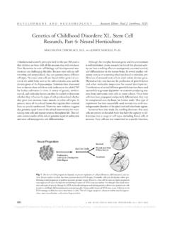
5 These will normally regress back into the peritoneal cavity by the end of the third month. (From Sadler TW, Langman s Medical Embryology, 5th edition, Williams & Wilkins, 1985, with permission.) The Umbilical Cord Harvey Kliman Sunday, October 29, 2006 Page 5 of 14 Figure 3. Fetus at ~53 days post-ovulation ( mm crown-rump length) showing distinct intestinal herniation into proximal Umbilical cord (arrow). Note twisting of Umbilical cord (arrow head). Figure 4. Remnants of the yolk sac stalk (A) and the allantois (B) can often be identified, especially near the fetal end of the cord. A) A cross section of yolk sac stalk (omphalomesenteric duct remnant) reveals a vacuolated, mucin rich epithelium, similar to normal intestinal epithelium. B) Cross section of an allantoic remnant reveals a flattened squamous epithelium similar to the urothelium found in the urogenital system.
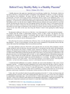
6 The Umbilical Cord Harvey Kliman Sunday, October 29, 2006 Page 6 of 14 The Umbilical cord normally contains two Umbilical arteries and one Umbilical vein. These are embedded within a loose, proteoglycan rich matrix known as Wharton s jelly (Figure 5). This jelly has physical properties much like a polyurethane pillow, which if you have ever tried twisting such a pillow you know is resistent to twisting and compression. This property serves to protect the critical vascular lifeline between the placenta and fetus (Figure 6). Figure 5. Cross section of normal Umbilical cord. Embedded within a spongy, proteoglycan rich matrix know as Wharton s jelly (W) are normally two arteries (A) and one vein (V). The Umbilical Cord Harvey Kliman Sunday, October 29, 2006 Page 7 of 14 Figure 6. The Umbilical cord protects the fetal vessels that connect the placenta and fetus.
7 A) Fetus and placenta from a 17 week gestation. B) Diagram of the circulation within the fetus, Umbilical cord and placenta. III. Abnormal Umbilical cord development Approximately 1% of all Umbilical cords contain only one artery rather than the normal two. Although many infants born with a single Umbilical artery have no obvious anomalies, single Umbilical artery has been associated with cardiovascular anomalies in 15-20% of such cases. While these anomalies could be the result of genetic factors alone, environmental factors may also play a part. For example, Naeye has shown an association between a single Umbilical artery and maternal smoking during pregnancy. As was stated previously, loops of bowel can be found within the proximal portion of the cord up until the end of the third month (Figures 2 and 3).
8 When this regression does not take place and herniation of peritoneal contents persists to term, a condition known as Meckel s diverticulum exists. Occasionally only a small portion of the vitelline duct may persist to term, leading to a vitelline cyst or fistula, which may need to be surgically removed after birth. The Umbilical Cord Harvey Kliman Sunday, October 29, 2006 Page 8 of 14 IV. Pathologic processes affecting the Umbilical cord As with any organ or tissue, the Umbilical cord can be subjected to both intrinsic and extrinsic pathological processes. Intrinsic processes include inflammation, knots and torsion, while extrinsic damage can occur iatrogenically following invasive, diagnostic procedures. The most common pathological finding in the Umbilical cord is funisitis (from the Latin cord inflammation ).
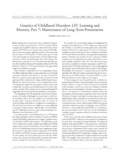
9 Funisitis is the result of neutrophils being chemotactically activated to migrate out of the fetal circulation towards the bacterially infected amnionic fluid (Figure 7). Since the ability of neutrophils to respond to chemokines and endotoxin is dependent on cellular maturation, it is not surprising to note that funisitis is only seen commonly after 20 weeks of gestation. Figure 7. Fetal neutrophil migration through the Umbilical cord (funisitis). A) In the presence of bacterial growth within the amnionic fluid (*), fetal neutrophils leave the Umbilical vessels (V) and migrate towards the amnionic cavity (arrow). In this case of severe funisitis, a wave of neutrophils and neutrophil breakdown products can be seen (arrow heads). B) Higher magnification of the edge of the neutrophil wave (arrow heads).
10 Less commonly, but with potentially devastating consequences, the Umbilical cord can become knotted (Figure 8). If the knot is loose, fetal circulation is maintained. However, if the knot is tightened, for example at the time of fetal descent through the birth canal, the tightening knot can occlude the circulation between the placenta and fetus, resulting in an intrauterine demise. The Wharton s jelly surrounding the fetal vessels is capable of withstanding significant torsional and compressional forces, as shown in Figure 9. Occasionally, however, Wharton s jelly does not develop in all portions of the cord. When this occurs, the fetal vessels are no longer protected from The Umbilical Cord Harvey Kliman Sunday, October 29, 2006 Page 9 of 14 torsional forces and they can become occluded if twisted sufficiently (Figure 10), again leading to an intrauterine demise.