Transcription of THORACIC OUTLET SYNDROME (T.O.S)
1 1. By Dr DG Harris THORACIC OUTLET SYNDROME ( ). WHAT IS THORACIC OUTLET SYNDROME ? The THORACIC OUTLET is the upper aperture of the chest, between the collar bone and the first narrow passageway is crowded with blood vessels that run out of the chest to the arm (subclavian vein and artery), as well as the nerves that exit the spine in the neck to supply the arm. The nerves fuse to form 3 large trunks (Brachial Plexus) and these run through the THORACIC OUTLET and split up again into separate nerves lower down.
2 THORACIC OUTLET SYNDROME refers to the symptoms that arise when these nerves or blood vessels are compressed at the THORACIC OUTLET . WHERE DOES THE COMPRESSION OCCUR, AND WHY? The THORACIC OUTLET is bordered by the collar bone at the top and front, the first rib below and two muscles, one in the front, and another behind. These muscles are called scalene muscles, and their function is to stabilise the first rib. They run from the spine, higher up and run downwards, and are attached to the first rib, one in the front and the other at the back of the rib.
3 The brachial plexus nerves and subclavian artery pass through the triangle formed between the first rib and scalene muscles. The subclavian vein lies in front of the first scalene muscle and behind the collar bone. Enlargement of the muscles, as well as scar tissue between them can compress the structures. The muscles may enlarge due to muscle imbalance following a shoulder injury or operation, and repetitive movements that exercise the muscle, such as certain sports, and certain work activities. Sport activities include swimming, ball throwing (cricket), and rugby.
4 Typical jobs that predispose to enlargement of these muscles are those where the arms are elevated a lot, such as mechanics, hairdressers, and schoolteachers (writing on blackboard). It is not uncommon in musicians. 2. Incorrect weight training may cause a muscle imbalance. A soft tissue injury (sprain) to the muscle can cause scar tissue. Congenital conditions such as abnormal bands that run over the nerves or between the muscles and an extra rib in the neck are other causes. Another area of compression is between the collar bone and the first rib.
5 Clavicle fractures may predispose to later scar formation, which may compress the structures later on. Fractures of the first rib may cause immediate damage to the brachial plexus, as it runs over the rib, making contact with it as it goes to the arm. If there is no immediate damage, symptoms may gradually develop later, as scar tissue grows around the rib, and encases the nerves and artery. Some people may have a very thick first rib, causing compression between it and the collar bone. The third area of compression may be further down, below the collar bone, where compression occurs by a thick pectoralis minor muscle where it attaches to the shoulder blade (scapula).
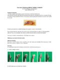
6 Poor posture and obesity may aggravate the condition. It occurs classically in females, with long necks, and drooping shoulders, as well as in stocky, muscular people. A painful shoulder following trauma or surgery may cause the arm to hang, and stretch the nerves over the rib if this is longstanding. WHAT ARE THE SYMPTOMS? Pressure on the blood vessels can reduce the blood flow to the arm and hand (especially in the elevated position) and cause them to tire easily, feel cold, and go pale. Pressure on the vein can cause the arm or hand to swell a bit.
7 Pressure on the nerves can cause vague, aching pains in the shoulder, neck, arm and hand. There may be a pain shooting all the way down to the fingers, and they may go numb. Headaches may occur. Overhead activities, carrying objects and driving may be affected. The arm may often go numb when lying on that side. The hand may feel clumsy and one may struggle with certain tasks, such as opening a jar, wringing out a cloth and there may be a tendency to drop things. 3. HOW IS THE DIAGNOSIS CONFIRMED? Diagnosis is confirmed by the typical symptoms, physical examination (to test clinical signs of artery and nerve compression) and some tests.
8 Unfortunately there is no one special test that can accurately exclude the problem of That is, a positive test can confirm it, however all the tests may be negative, but the patient may still have , and may suffer for a long time before the diagnosis has been made. Some patients may have had neck or shoulder surgery before TOS is diagnosed. In a lot of cases TOS may ultimately be diagnosed by the surgical decompression operation ( open up and see ). TOS is often only diagnosed after excluding other conditions that may be present with similar symptoms, and these need to be excluded.
9 These are shoulder problems ( rotator cuff injury), neck problems (prolapsed disc), ulnar nerve entrapment at the elbow & carpal tunnel entrapment at the wrist. Rarely, a cancerous growth at the top of the lung may invade the brachial plexus. (Usually in patients over 55 years). The following investigations are routinely performed: o Evaluation by a shoulder surgeon to exclude shoulder pathology, with clinical examination, ultrasound and x-rays and possibly MRI scan of the shoulder. o X-rays of the cervical spine to assess for cervical ribs and check the alignment, and if indicated, MRI scan of the neck.
10 O Chest X-Ray to check the lung, and look for the clavicle and first rib deformities. o Other tests performed, include: o Nerve conduction tests (done by neurologists) together with neurological evaluation. These may on occasion be of value, but a negative test does not mean that TOS is not present. The test is expensive. o Multi-slice CT scan (CT Angiogram). This can be performed with the arms at the side and then with the arms elevated above the head, and can confirm dynamic compression of the artery. This may be helpful, but may not diagnose nerve compression.